





Foraminal stenosis: symptoms, MRI findings and treatment decisions
Foraminal stenosis narrows the opening through which a spinal nerve exits. It may cause radicular pain, tingling, numbness or weakness, but imaging severity does not always match symptoms.
Diagnosis depends on correlation between the level, side, symptom distribution, neurological findings and functional impact.
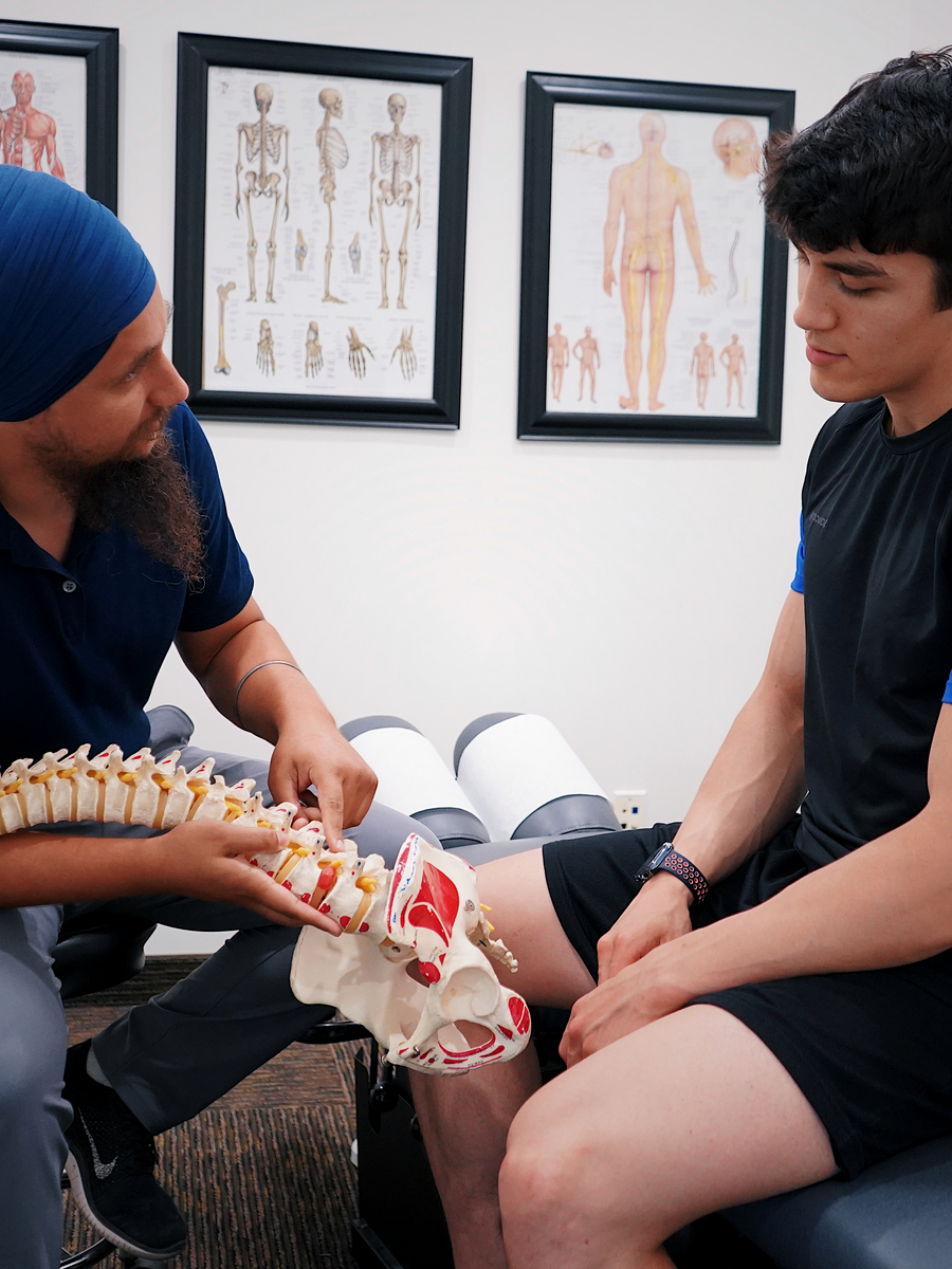
How it may feel
Symptoms often follow the exiting nerve root and may be provoked by standing, walking or extension. Hip disease, peripheral neuropathy and central stenosis may overlap.
Neurological testing examines the muscles, sensation and reflexes of the suspected root.
| Feature | Possible meaning | Caution |
|---|---|---|
| Radicular pain | Irritation of the exiting root or ganglion. | Pain maps overlap. |
| Numbness or tingling | Sensory dysfunction in a root distribution. | Peripheral entrapment may mimic it. |
| Weakness | Motor involvement. | Progressive weakness increases urgency. |

MRI and clinical correlation
MRI assesses perineural fat, root deformation and disc or facet changes. Grading can be reproducible, but correlation with pain and disability is imperfect.
Assessment includes walking, extension tolerance, strength, sensation, reflexes and positional effects.
Level and side
The abnormal foramen should match the symptomatic limb.
Functional provocation
Standing or walking may reproduce symptoms.
Differential diagnosis
Central stenosis, hip disease and neuropathy may coexist.

Conservative, interventional and surgical options
Initial care may include education, activity modification, exercise and medication when appropriate. Strength and function should be monitored.
Injections may be discussed. Surgery such as foraminotomy is considered when symptoms and imaging correlate, conservative care fails or deficits progress.
- Identify positions that provoke or relieve symptoms.
- Build walking and strength tolerance carefully.
- Review the entire MRI because several levels may look abnormal.
- Consider stability before decompression or fusion.
Choosing the next step
The decision is influenced by severity, deficit, duration, function, stability and confidence in anatomical correlation.
Severe fixed stenosis may require a different strategy than a monitored non-surgical trial.
| Situation | Reasonable discussion | Key question |
|---|---|---|
| Mild stable symptoms | Conservative care and monitoring. | Can function improve safely? |
| Persistent pain with concordant MRI | Targeted non-surgical or interventional options. | Is measurable function improving? |
| Progressive weakness or severe refractory symptoms | Prompt surgical opinion. | Is direct foraminal decompression required? |

Questions to ask before choosing care
Before beginning care for foraminal stenosis, ask the provider to explain the working diagnosis and the findings that support it. The discussion should distinguish a structural description from the clinical syndrome, clarify whether neurological function is stable and identify the specific outcome the treatment is intended to improve.
A high-quality plan also explains the expected timeline, reasonable alternatives, possible harms, cost and the criteria for changing course. No treatment should continue indefinitely because it produced a brief change in pain. The decision should be reviewed against measurable function and safety.
What is the target?
Ask which structure, mechanism or functional limitation the treatment is intended to address.
What result is realistic?
Clarify the expected change in pain distribution, walking, sleep, work or daily activity.
What are the alternatives?
Compare education, exercise, medication, procedures, technology-assisted care and surgery when relevant.
When will the plan be reviewed?
Set an early review point and define the findings that would stop, modify or escalate care.
How to monitor progress
Progress should be tracked with more than a pain score. Useful measures include how far symptoms travel, strength, sensation, walking tolerance, sleep, medication use and the ability to perform meaningful activities. Neurological change should always be documented separately from pain relief.
Temporary fluctuations are common. The important question is whether the overall trajectory is safer and more functional. New weakness, expanding numbness, altered bladder or bowel function or a substantial loss of walking ability should trigger reassessment rather than automatic continuation of the same treatment.
Continue with specialized resources
The Spine Page is an educational blog. The links below lead to independent specialized websites.
Clinique TAGMED — spinal decompression
Service-specific information about motorized spinal decompression and disc-related conditions.
SOS Sciatique — nerve-root symptoms
Focused guides about sciatica, warning signs and treatment questions.
Frequently asked questions
Is it the same as central stenosis?
No.
Can it occur at L5–S1?
Yes.
Does severe MRI narrowing always cause severe pain?
No.
Can standing worsen symptoms?
Yes.
Can it cause foot drop?
Severe L5 dysfunction may contribute.
Is an injection diagnostic?
It may assist, but is not perfectly specific.
Can exercise assist?
Yes, when adapted.
Can decompression assist?
Suitability varies.
When is surgery considered?
When correlation is strong and symptoms or deficits persist.
Can The Spine Page grade my MRI?
No.
Sources consulted
- Sundaram et al. — Assessment of Lumbar Foraminal Stenosis
- Katz et al. — Diagnosis and Management of Lumbar Spinal Stenosis
- American College of Radiology — Appropriateness Criteria: Low Back Pain
Last editorial review: July 2026. This page provides general education and does not replace individualized medical assessment.
Editorial review : Dr. Sylvain Desforges, B.Sc., D.O., N.D., osteopath — The Spine Page
Explore radiculopathy and decompression
Continue with L4–L5–S1 patterns or review spinal decompression.
The Spine Page — The best treatments for your spinal problems — www.thespinepage.com
Topic index: Spinal and foraminal stenosis
Explore this topic cluster by search intent. Each link leads to a distinct guide directly related to the subject.