





Sacroiliitis vs sacroiliac joint dysfunction: inflammatory or mechanical pain?
Sacroiliitis is inflammation of one or both sacroiliac joints and may be associated with axial spondyloarthritis, infection or another inflammatory disorder. Sacroiliac joint dysfunction usually refers to non-inflammatory mechanical pain around the joint.
Both conditions can cause buttock, posterior pelvic, groin or referred thigh pain. Mechanical SI-joint pain is often linked to trauma, pregnancy, altered load transfer, adjacent fusion, leg-length asymmetry or degenerative change. Inflammatory sacroiliitis more often begins young, improves with activity, causes night pain and morning stiffness and may coexist with psoriasis, uveitis, enthesitis or inflammatory bowel disease. Infection is a separate urgent form of sacroiliitis.

Assessment and differential diagnosis
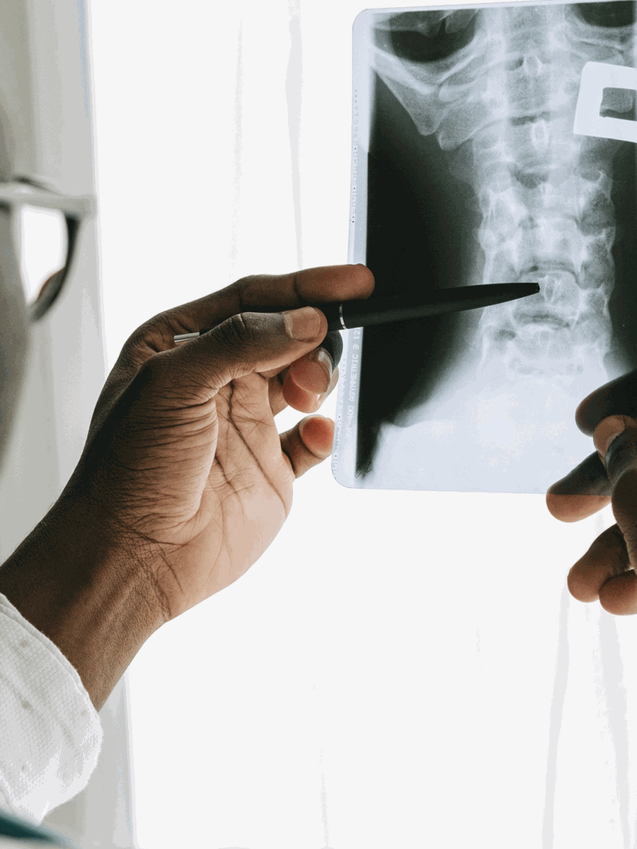
Mechanical SI-joint pain is assessed with history, lumbar and hip examination and a cluster of pain-provocation tests; three or more positive provocation tests increase suspicion but are not perfect. Image-guided intra-articular anaesthetic block can improve diagnostic confidence before an invasive procedure. Inflammatory assessment considers age at onset, night pain, morning stiffness, exercise response, family history and extra-articular disease. Pelvic radiographs evaluate structural sacroiliitis, while MRI using T1 and fluid-sensitive sequences detects active inflammation and structural lesions when radiographs are negative. HLA-B27 and inflammatory markers can support but do not confirm or exclude the diagnosis.
| Clinical pattern | Possible next step | Why it matters |
|---|---|---|
| Pain after trauma, pregnancy or loading with positive provocation cluster | Mechanical SI-joint dysfunction more likely | Conservative mechanical care is usually first line. |
| Onset before 45 with night pain, stiffness and inflammatory features | Inflammatory sacroiliitis possible | Rheumatology referral should be considered. |
| Fever, severe unilateral SI pain or immunosuppression | Infectious sacroiliitis possible | Urgent blood tests and MRI are required. |
| Imaging abnormality without concordant symptoms | Do not assume the SI joint is the pain source | Clinical correlation remains essential. |

Treatment and decision-making
Mechanical SI-joint dysfunction is initially managed with education, activity modification, pelvic and trunk rehabilitation, selected manual therapy and medication after risk assessment. A pelvic belt may help selected postpartum patients. Image-guided corticosteroid injection, lateral-branch radiofrequency treatment or minimally invasive fusion may be considered for persistent confirmed mechanical pain after non-surgical care. Inflammatory sacroiliitis requires rheumatology-led treatment with exercise, NSAIDs when appropriate and disease-modifying or biological therapy when indicated. Infectious sacroiliitis requires antimicrobial treatment and sometimes drainage.
How to interpret the condition in practice
The words sacroiliitis and SI-joint dysfunction should not be used as synonyms. One implies inflammation that may require rheumatology or infection treatment; the other usually describes mechanical pain and requires confirmation that the joint is actually the pain source.
Ask when it began
Young onset raises inflammatory suspicion.
Check night and morning symptoms
Inflammatory pain often has a different daily pattern.
Use provocation clusters
One positive mechanical test is insufficient.
Image for the right question
MRI inflammation and mechanical dysfunction are different targets.
Review extra-articular disease
Psoriasis, uveitis and bowel disease matter.
Do not inject blindly
A precise image-guided procedure is preferable when an injection is used.
How to monitor the condition and avoid interpretation errors
A credible plan should define what improvement means before treatment begins and should explain how the diagnosis will be reconsidered if the expected response does not occur. Pain intensity can change independently from neurological function, walking tolerance, sleep or participation. For uncommon conditions, progression and safety are often more important than day-to-day symptom fluctuation.
Create a baseline
Record symptom distribution, strength, sensation, walking, sleep, medication use and the activities that matter most.
Use a planned review point
Decide when the response will be reviewed rather than continuing automatically until a treatment package is completed.
Separate relief from diagnosis
Temporary improvement after medication, injection, manual care or a device does not prove the proposed structure was the true cause.
Watch the neurological trajectory
New weakness, spreading numbness, gait change or autonomic symptoms matter even if pain decreases.
Review alternative causes
Degenerative, inflammatory, infectious, oncological and postoperative mechanisms may overlap.
Escalate proportionately
Imaging, specialist referral or surgery should be considered when the risk or functional loss justifies it.
Common errors to avoid
- Treating the most unusual imaging finding instead of the clinically relevant lesion.
- Assuming that a stable imaging abnormality explains a changing symptom pattern.
- Using one negative test to exclude a condition that remains clinically plausible.
- Repeating passive treatment despite no measurable functional progress.
- Generalizing results from a small specialist series to every person with the same label.
- Delaying urgent assessment to complete a predetermined number of visits.
Shared decision-making is strongest when uncertainty is stated directly. A provider should explain what is confirmed, what remains probable, which alternatives still matter and what findings would change the plan. For rare or controversial diagnoses, a second opinion from a clinician experienced with the specific condition may be more useful than adding another non-specific treatment.
Questions before choosing care
What is the leading diagnosis?
Ask which symptoms and examination findings support it.
What alternative remains possible?
Common competing causes should be reviewed before an invasive procedure.
What test changes management?
Avoid testing or imaging that cannot alter the next step.
What outcome matters?
Track function, neurological safety and meaningful activity.
A single symptom, image or treatment response rarely establishes the complete cause by itself.
Relevant specialized resources
Some resources may share ownership or editorial direction; this relationship is disclosed and the links are included for contextual relevance.
Clinique TAGMED — osteopathy
Clinical information about selected mechanical pelvic and spinal care.
Frequently asked questions
Is sacroiliitis the same as SI-joint dysfunction?
No.
Can mechanical SI pain have a normal MRI?
Yes.
Can inflammatory sacroiliitis have a normal X-ray?
Yes, especially early.
How many provocation tests are useful?
A cluster of three or more positive tests increases suspicion.
Does HLA-B27 confirm the diagnosis?
No.
Can pregnancy cause SI-joint dysfunction?
Yes.
Can psoriasis be relevant?
Yes, for inflammatory spondyloarthritis.
What is a diagnostic SI-joint block?
An image-guided anaesthetic injection used to test whether the joint is the pain source.
When is fusion considered?
Only after persistent confirmed mechanical pain fails appropriate non-surgical care.
Does The Spine Page diagnose sacroiliitis?
No.
Sources consulted
- Waldman et al. — SI-joint dysfunction differential diagnosis and treatment
- NICE — Spondyloarthritis diagnosis and management
- ACR Appropriateness Criteria — Inflammatory Back Pain
- Polly — SI joint state-of-the-art review
Last editorial review: July 2026. General educational information only.
Dr. Sylvain Desforges, B.Sc., D.O., N.D., osteopath — The Spine Page
Continue exploring spine health
Use the condition and treatment hubs to compare related guides.
The Spine Page — The best treatments for your spinal problems — www.thespinepage.com