





CT scan vs MRI for the spine: what each test shows and when it is used
CT and MRI answer different spinal questions. CT excels at bone and fracture detail, while MRI provides superior information about discs, nerves, spinal cord, marrow and many soft tissues.
The best test is not necessarily the most detailed one. Imaging should be selected only when the result is likely to change diagnosis, treatment or urgency. Unnecessary imaging can reveal incidental findings that increase concern without improving care.

Assessment and differential diagnosis
The clinical question determines the test. Suspected fracture, complex bone anatomy or hardware may favor CT. Suspected disc herniation, cord compression, infection, cancer or nerve-root compromise often favors MRI. Contrast is used selectively rather than automatically.
| Clinical situation | Possible next step | Why it matters |
|---|---|---|
| Acute fracture or bone detail | CT often preferred | Fast and excellent for osseous anatomy. |
| Radiculopathy or myelopathy | MRI often preferred | Neural structures and soft tissues are assessed. |
| Routine uncomplicated back pain | Neither may be needed initially | Imaging should change management. |
Treatment and management
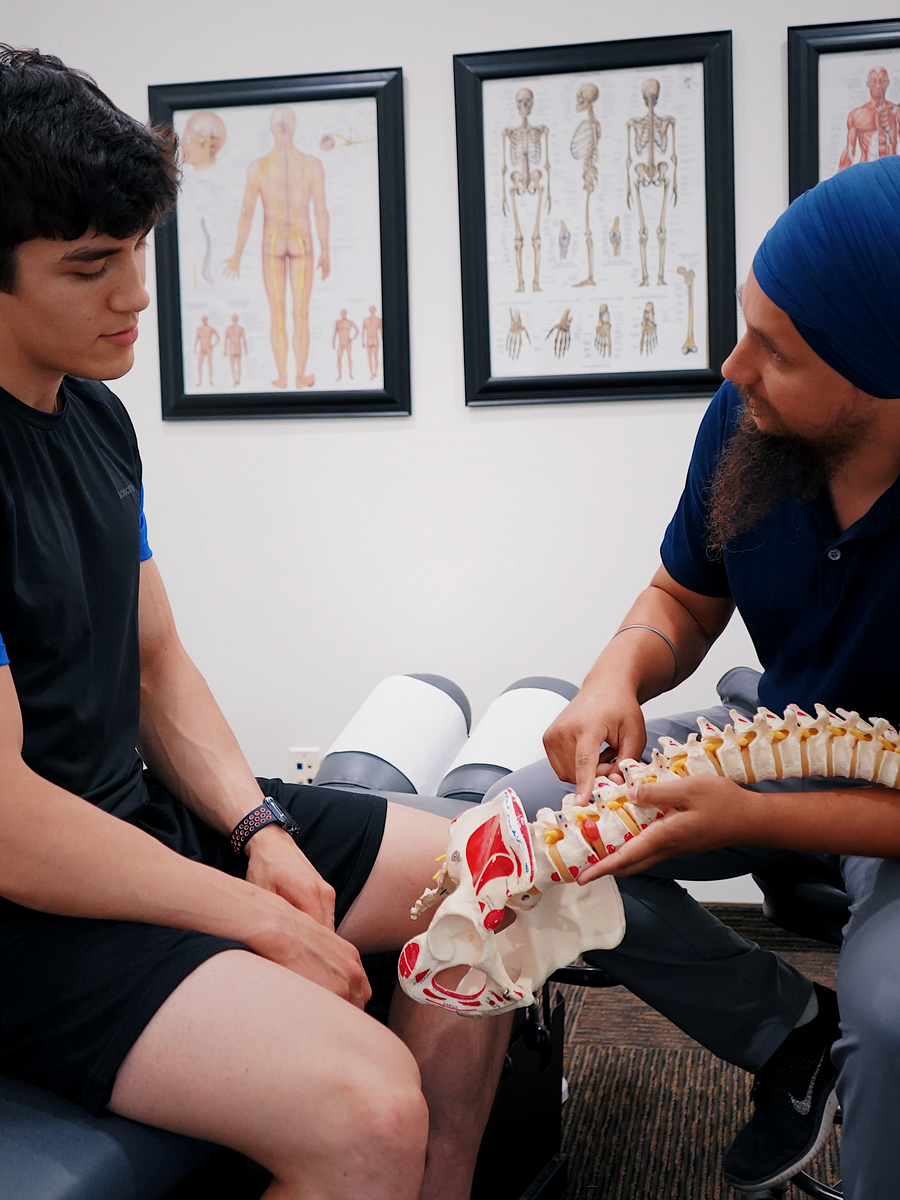
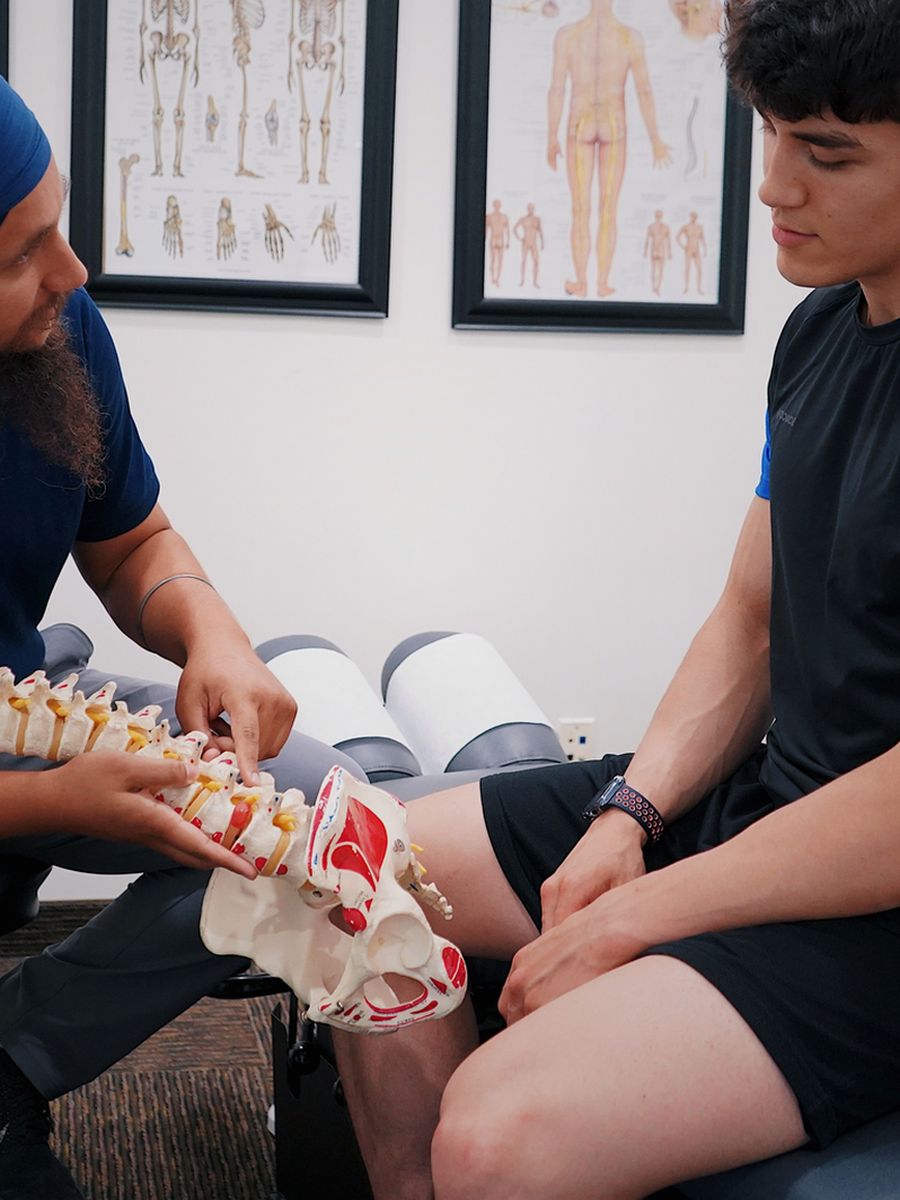
Imaging results should be translated into a clinical plan. A report describing degeneration, bulging or stenosis does not establish the source of symptoms by itself. The provider should explain which finding is relevant and what decision it changes.
Questions for shared decision-making
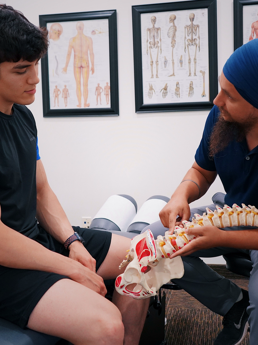
Before choosing care, ask the provider to explain the working diagnosis, expected outcome, alternatives and review point.
What is the target?
Identify the structure, mechanism or functional problem being addressed.
What outcome matters?
Define measurable goals for pain, walking, sleep, work or daily activity.
What are the alternatives?
Compare active care, medication, procedures, technology and surgery when relevant.
When should the plan change?
Set criteria for stopping, modifying or escalating care.
Related guides
When is MRI needed?
EMG and nerve-conduction studies
Specialized external resources
These links lead to independent specialized websites and do not change the educational role of The Spine Page.
Clinique TAGMED
Clinical information about motorized spinal decompression and disc-related conditions.
SOS Sciatique
Focused guides about sciatica, nerve-root symptoms and warning signs.
Frequently asked questions
Which is better, CT or MRI?
Neither is universally better; they answer different questions.
Does CT show a herniated disc?
It may, but MRI is usually better for discs and nerves.
Does MRI show fractures?
Yes, and it can show fracture acuity and marrow change.
Does MRI use radiation?
No.
Is contrast always needed?
No.
Can I have MRI with an implant?
It depends on the implant and MRI safety information.
Which is faster?
CT is usually faster.
Is imaging needed for every back pain episode?
No.
Can imaging findings be incidental?
Yes.
Does The Spine Page order imaging?
No.
Sources consulted
- American College of Radiology — Appropriateness Criteria
- NICE — Low back pain and sciatica recommendations
- American College of Radiology — Thoracic Back Pain
Last editorial review: July 2026. General educational information only.
Dr. Sylvain Desforges, B.Sc., D.O., N.D., osteopath — The Spine Page
Continue exploring spine health
Use the condition and treatment hubs to compare the next steps.
The Spine Page — The best treatments for your spinal problems — www.thespinepage.com
Topic index: Imaging and diagnostic tests
Explore this topic cluster by search intent. Each link leads to a distinct guide directly related to the subject.