





When is an MRI needed for back pain or sciatica?
Most uncomplicated acute low back pain and radiculopathy does not require immediate imaging. MRI becomes useful when serious pathology is suspected, deficits are present or persistent symptoms make intervention realistic.
The better question is not whether MRI can show something, but whether the result is likely to change management in a beneficial way.
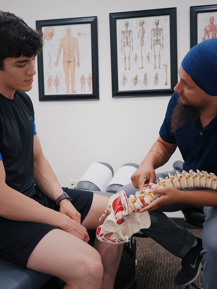
When MRI becomes more relevant
MRI is valuable when it can identify neural compression, infection, malignancy, fracture complication or postoperative pathology and influence treatment.
Contrast is not required for every case; prior surgery, infection or tumour may change the protocol.
| Clinical scenario | Imaging relevance | Reason |
|---|---|---|
| Persistent radiculopathy after care | MRI often appropriate if intervention is considered. | Defines the level and type of compression. |
| Suspected cauda equina | Urgent MRI. | Assesses severe compression of several roots. |
| Cancer or infection concern | MRI, sometimes with contrast. | Evaluates destructive or inflammatory pathology. |

What should happen before ordering MRI?
The clinician should document symptoms, red flags, neurological examination, prior care and the decision that might follow the scan.
A normal MRI does not invalidate pain, and an abnormal MRI does not automatically prove the cause.
Clinical question
Specify what condition is being investigated.
Expected action
Decide what changes with a positive or negative result.
Protocol
Choose region and contrast according to the question.

What happens after MRI?
Images should be correlated with the side and level of symptoms and neurological findings. The report should distinguish common ageing from important compression.
If MRI does not identify a concordant target, highly specific invasive treatment at an incidental level may not be appropriate.
- Bring previous imaging when relevant.
- Ask whether the finding explains the exact pattern.
- Clarify common versus important findings.
- Use the result to confirm or revise the plan.
A practical MRI framework
The table summarizes broad principles; actual decisions depend on the full situation.
Emergency access and protocols vary by institution.
| Question | If no | If yes |
|---|---|---|
| Are red flags or progressive deficits present? | Conservative care may continue. | Urgent or expedited imaging may be appropriate. |
| Have symptoms persisted despite care? | Continue monitoring if improving. | MRI may assist before intervention. |
| Will the result change treatment? | Routine imaging adds less value. | Order the appropriate protocol. |
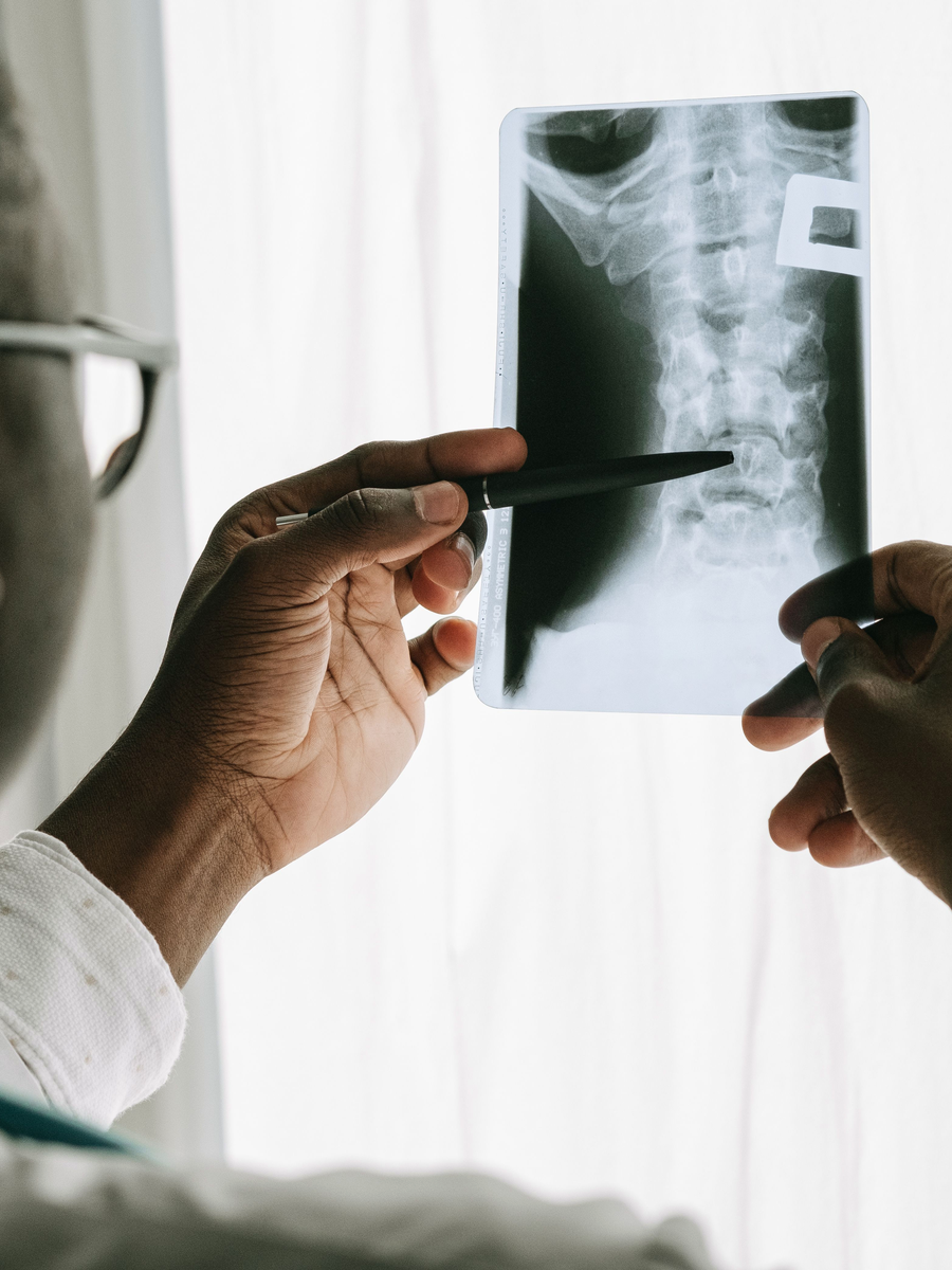
Questions to ask before choosing care
Before beginning care for when is an mri needed for back pain or sciatica?, ask the provider to explain the working diagnosis and the findings that support it. The discussion should distinguish a structural description from the clinical syndrome, clarify whether neurological function is stable and identify the specific outcome the treatment is intended to improve.
A high-quality plan also explains the expected timeline, reasonable alternatives, possible harms, cost and the criteria for changing course. No treatment should continue indefinitely because it produced a brief change in pain. The decision should be reviewed against measurable function and safety.
What is the target?
Ask which structure, mechanism or functional limitation the treatment is intended to address.
What result is realistic?
Clarify the expected change in pain distribution, walking, sleep, work or daily activity.
What are the alternatives?
Compare education, exercise, medication, procedures, technology-assisted care and surgery when relevant.
When will the plan be reviewed?
Set an early review point and define the findings that would stop, modify or escalate care.
How to monitor progress
Progress should be tracked with more than a pain score. Useful measures include how far symptoms travel, strength, sensation, walking tolerance, sleep, medication use and the ability to perform meaningful activities. Neurological change should always be documented separately from pain relief.
Temporary fluctuations are common. The important question is whether the overall trajectory is safer and more functional. New weakness, expanding numbness, altered bladder or bowel function or a substantial loss of walking ability should trigger reassessment rather than automatic continuation of the same treatment.
Continue with specialized resources
The Spine Page is an educational blog. The links below lead to independent specialized websites.
Clinique TAGMED — spinal decompression
Service-specific information about motorized spinal decompression and disc-related conditions.
SOS Sciatique — nerve-root symptoms
Focused guides about sciatica, warning signs and treatment questions.
Frequently asked questions
Do I need MRI for every episode?
No.
Can X-ray show a herniated disc?
No.
When is MRI urgent?
With cauda equina, major progressive deficit or serious pathology.
Why is contrast used?
For selected infection, tumour or postoperative questions.
Can MRI identify the exact pain source?
Not always.
What if MRI is normal?
Pain can still be real.
What if several abnormalities appear?
The clinician decides which, if any, match symptoms.
Is MRI harmful?
It has no ionizing radiation but has implant and incidental-finding considerations.
Can I request MRI immediately?
You can discuss it, but routine early imaging is often discouraged.
Does The Spine Page order imaging?
No.
Sources consulted
- American College of Radiology — Appropriateness Criteria: Low Back Pain
- NICE — Low back pain and sciatica in over 16s
- Brinjikji et al. — Imaging features of spinal degeneration in asymptomatic populations
Last editorial review: July 2026. This page provides general education and does not replace individualized medical assessment.
Editorial review : Dr. Sylvain Desforges, B.Sc., D.O., N.D., osteopath — The Spine Page
Use imaging in the right pathway
Review warning signs or browse the condition guides.
The Spine Page — The best treatments for your spinal problems — www.thespinepage.com