





Schmorl nodes on MRI: incidental endplate defect or source of back pain?
A Schmorl node is disc material that has pushed vertically through a vertebral endplate into the adjacent vertebral body. These nodes are common and usually incidental, but an acute inflamed node with surrounding marrow oedema can sometimes produce focal back pain.
Schmorl nodes occur most often near the thoracolumbar junction and upper lumbar spine. They may result from developmental endplate weakness, axial loading, degeneration, trauma, Scheuermann disease, osteoporosis or other conditions that weaken the endplate. The imaging finding alone does not establish pain, injury timing or the need for treatment.

Assessment and differential diagnosis
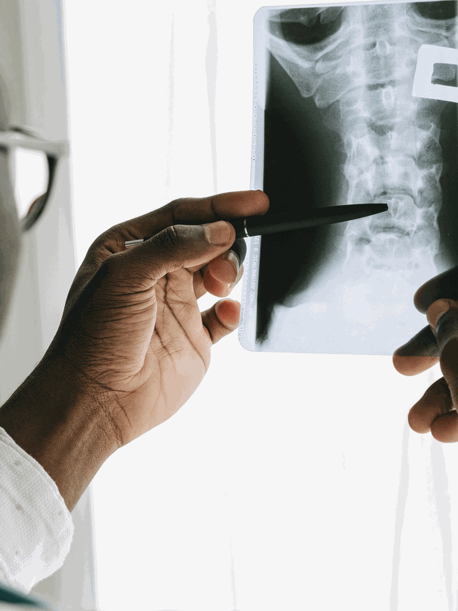
MRI shows the endplate defect and surrounding marrow signal. An old inactive node usually has a well-defined sclerotic margin and no oedema. A potentially acute or symptomatic node may show low T1 and high T2 or STIR marrow signal, enhancement and local tenderness, but these findings are still not perfectly specific. CT can better show cortical detail and sclerosis. The differential diagnosis includes compression fracture, infection, metastasis, inflammatory endplate disease and Modic change. Clinical history, laboratory findings and follow-up imaging may be necessary when the appearance is atypical.
| Clinical pattern | Possible next step | Why it matters |
|---|---|---|
| Old incidental node without marrow oedema | No node-specific treatment | The finding is usually clinically inactive. |
| Acute focal pain with oedematous node and no serious alternative diagnosis | Conservative care and follow-up | Inflammation may settle over time. |
| Atypical destructive lesion or systemic symptoms | Investigate infection, tumour or fracture | Do not assume a benign Schmorl node. |
| Node with disc herniation or neurological compression | Treat the clinically relevant lesion | The node may be secondary context. |

Treatment and decision-making
Most Schmorl nodes require no treatment. A suspected acute symptomatic node is generally managed with temporary activity modification, analgesia after risk assessment, gradual rehabilitation and treatment of bone-health or loading factors. Bracing, injections, vertebral augmentation or fusion have been described in rare refractory cases, but evidence is limited to case reports and small series. The goal is not to erase the MRI finding but to restore function while excluding fracture, infection and tumour.
How to interpret the finding in practice
The key distinction is between a common old endplate defect and a new active lesion. The radiology report should be interpreted with symptom onset, bone health, trauma history and the surrounding marrow signal.
Review the marrow
Oedema and enhancement may indicate recent activity.
Check the cortex
CT can clarify fracture and sclerosis.
Assess bone health
Osteoporosis and metabolic disease can weaken endplates.
Exclude systemic disease
Fever, cancer and inflammatory markers change the differential.
Avoid overdiagnosis
A node without matching symptoms should not drive treatment.
Use follow-up wisely
Resolution of oedema may support healing when the diagnosis was uncertain.
How to monitor the condition and avoid interpretation errors
A diagnosis based partly on imaging should be reviewed over time rather than treated as a permanent explanation for every symptom. The condition may remain stable, become less clinically relevant or coexist with a second problem. A useful follow-up records the neurological examination, the activities that are limited, the treatment response and the circumstances that would justify new imaging or specialist referral.
Record a functional baseline
Document walking, sitting, lifting, sleep, work, sport and self-care before beginning treatment.
Separate local and neurological symptoms
Local pain, referred pain, numbness and objective weakness should not be treated as interchangeable outcomes.
Use imaging for a question
Repeat imaging is most useful when the result may alter diagnosis, urgency or treatment.
Review competing diagnoses
Hip, sacroiliac, vascular, inflammatory, peripheral nerve and persistent-pain mechanisms may coexist.
Define a stopping rule
A procedure or treatment package should not continue automatically without meaningful progress.
Communicate uncertainty
A credible explanation distinguishes what is confirmed, what is probable and what remains unknown.
Common interpretation errors
- Assuming that the largest or most unusual MRI finding must be the pain source.
- Using temporary symptom relief as proof of a structural diagnosis.
- Ignoring neurological change because pain intensity has decreased.
- Repeating passive treatment when function and participation are not improving.
- Applying the results of a small specialist case series to every person with the same imaging label.
- Delaying medical or surgical referral to complete a predetermined number of visits.
Shared decision-making is strongest when the provider explains the expected natural history, the quality of the evidence, the main alternatives and the risks of both treatment and observation. The person should know which changes can be monitored routinely and which require prompt reassessment. For uncommon or controversial diagnoses, a second opinion from a clinician experienced with the specific condition may be more valuable than adding another non-specific treatment.
Questions before choosing care
What is the leading diagnosis?
Ask which symptoms and examination findings support it.
What alternative remains possible?
Common competing causes should be reviewed before an invasive procedure.
What test changes management?
Avoid imaging or procedures that cannot alter the next step.
What outcome matters?
Track function, neurological safety and meaningful activity.
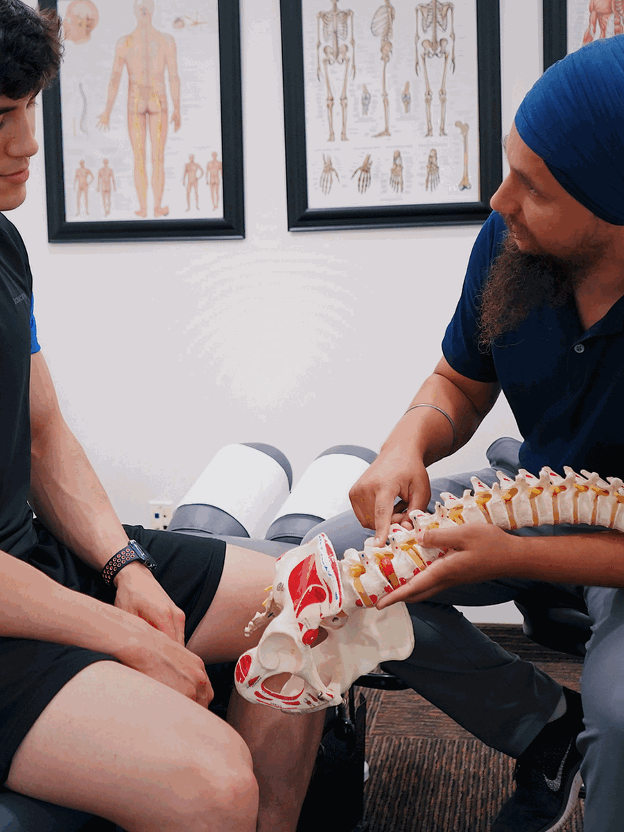
A single image or label rarely establishes the complete cause or the best treatment by itself.
Relevant specialized resources
Some resources may share ownership or editorial direction; this relationship is disclosed and the links are included for contextual relevance.
Frequently asked questions
Is a Schmorl node a herniated disc?
It is a vertical intravertebral disc herniation rather than the usual posterior or foraminal herniation.
Are Schmorl nodes common?
Yes.
Do they always cause pain?
No.
What suggests an active painful node?
Recent focal pain with surrounding marrow oedema may increase suspicion.
Can trauma cause a node?
Trauma or repetitive axial loading may contribute.
Are they related to Scheuermann disease?
Multiple endplate defects and Schmorl nodes can occur in Scheuermann disease.
Can a node look like cancer or infection?
Atypical acute lesions may require differential investigation.
Does the node need surgery?
Almost never; surgery is reserved for exceptional refractory cases.
Can the node disappear?
The defect may remain while inflammation and symptoms resolve.
Does The Spine Page diagnose Schmorl nodes?
No.
Sources consulted
- Zhang et al. — Critical review of Schmorl nodes
- Mok et al. — Population-based MRI study
- Samartzis et al. — MRI classification and disc degeneration
- Takahashi et al. — Symptomatic vs asymptomatic Schmorl nodes
Last editorial review: July 2026. General educational information only.
Dr. Sylvain Desforges, B.Sc., D.O., N.D., osteopath — The Spine Page
Continue exploring spine health
Use the condition and treatment hubs to compare related guides.
The Spine Page — The best treatments for your spinal problems — www.thespinepage.com