





Cervical spondylosis vs cervical disc herniation: neck and arm symptoms
Cervical spondylosis describes age-related disc, facet, ligament and bone changes across one or more levels. A cervical disc herniation is a more specific displacement of disc material. Either can cause neck pain, radiculopathy or myelopathy.
Spondylosis is a broad degenerative process that may include disc-height loss, osteophytes, facet arthritis, uncovertebral enlargement and ligament thickening. A herniation may be soft, focal and relatively acute or may coexist with chronic spondylosis. The label does not reveal which structure is symptomatic. The clinically important question is whether there is local mechanical pain, nerve-root compression or spinal-cord dysfunction.

Assessment and differential diagnosis
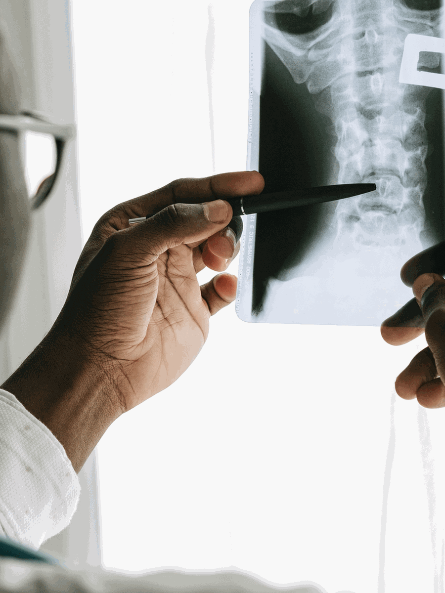
History reviews onset, trauma, neck and arm distribution, hand function, balance and symptom progression. Examination includes strength, sensation, reflexes, gait, dexterity and upper-motor-neuron signs. ACR criteria indicate that routine MRI is not automatically appropriate for uncomplicated acute neck pain without red flags, while MRI becomes more relevant for chronic radiculopathy, progressive neurological findings or suspected myelopathy. CT can define osteophytes and bone anatomy, and flexion–extension radiographs may be used selectively when instability is suspected.
| Clinical pattern | Possible next step | Why it matters |
|---|---|---|
| Multilevel disc-height loss and osteophytes with stiffness | Cervical spondylosis likely contributes | Treat the symptomatic mechanism rather than every level. |
| Focal soft-disc lesion with matching arm symptoms | Disc herniation may be dominant | Monitor motor and sensory function. |
| Cord compression with gait or hand dysfunction | Cervical myelopathy pathway | Prompt specialist assessment is appropriate. |
| Imaging changes without concordant symptoms | Observation and alternative diagnosis review | Age-related findings are common. |
Treatment and decision-making
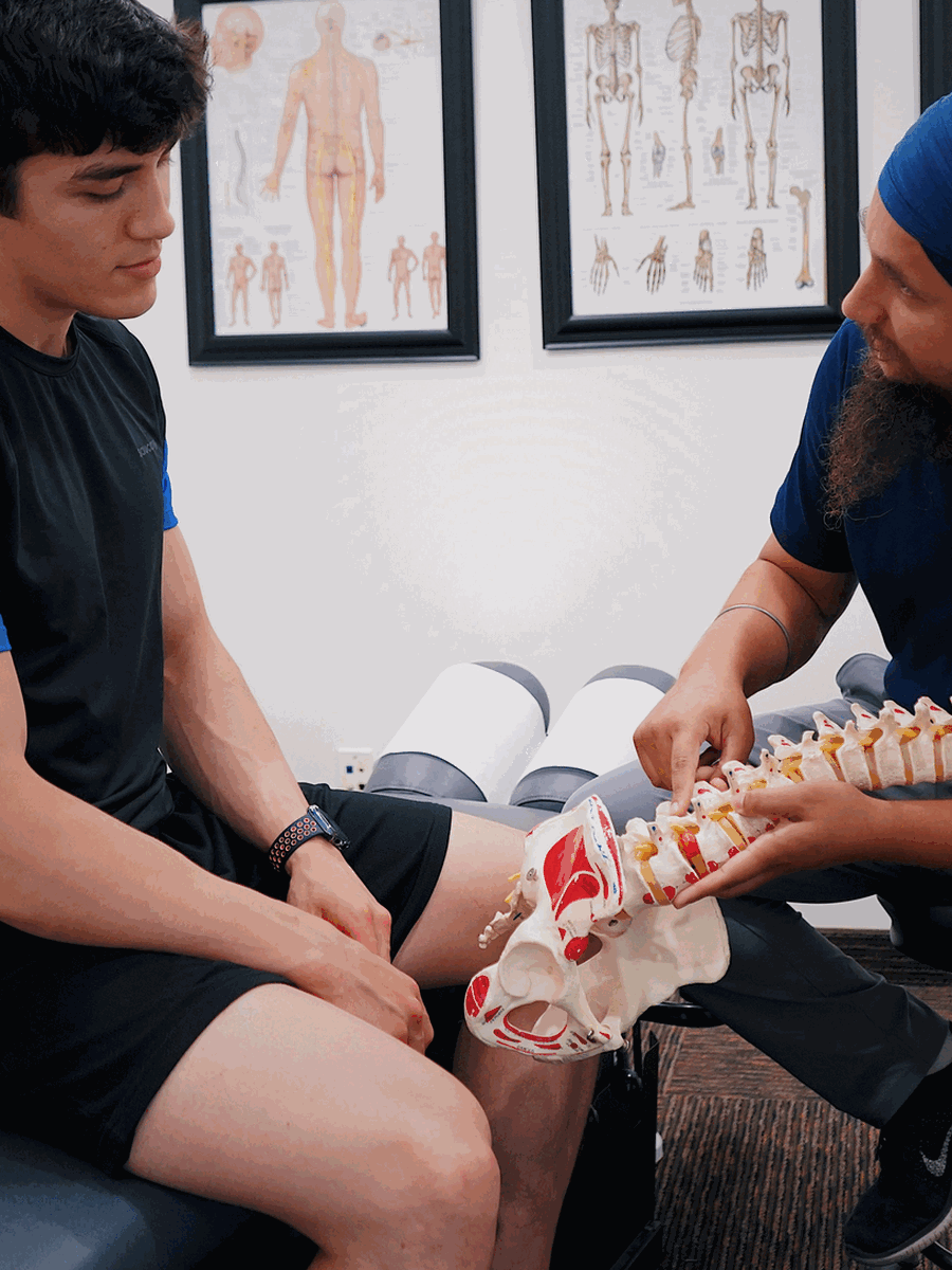
Stable neck pain or radiculopathy may begin with education, activity modification, exercise, medication and selected manual or physical therapies. Epidural injection may be considered for selected radicular pain after risk review. Surgery is considered when myelopathy, progressive weakness or severe persistent concordant radiculopathy is present. Anterior discectomy and fusion, disc replacement, posterior foraminotomy and laminoplasty are different operations chosen according to the lesion, levels, alignment and neurological objective.
How to interpret the finding in practice
The terms are not mutually exclusive. A person can have a focal herniation superimposed on multilevel spondylosis, and the treatment target may be only one level or one neurological structure.
Separate local and arm pain
Axial pain and radiculopathy may have different generators.
Look for cord signs
Gait and hand changes are more urgent than stiffness alone.
Identify soft versus hard compression
Disc material and osteophytes behave differently.
Count the levels
Single-level and multilevel surgery have different implications.
Assess alignment
Kyphosis or instability changes surgical planning.
Avoid age-based assumptions
Degeneration can be asymptomatic.
How to monitor progress and avoid treatment errors
A credible treatment plan should define what improvement means before care begins. Pain intensity alone is not enough, because pain may fluctuate while neurological function, walking, sleep or daily activity changes in another direction. The diagnosis should also be reconsidered when the expected response does not occur.
Establish a baseline
Record symptom distribution, strength, sensation, walking, sleep, medication use and the activities that matter most.
Use a planned review point
Decide when progress will be evaluated rather than continuing automatically until a package is completed.
Separate relief from diagnosis
Temporary improvement after an injection, manual treatment or device does not prove that the proposed structure was the true cause.
Watch the neurological trajectory
New weakness, spreading numbness or balance change matters even if pain has decreased.
Revisit competing diagnoses
Hip, sacroiliac, vascular, inflammatory and postoperative causes may overlap with the initial explanation.
Escalate proportionately
Imaging, specialist referral or surgery should be considered when the risk or functional loss justifies it.
Common mistakes to avoid
- Treating the largest imaging abnormality rather than the clinically relevant lesion.
- Assuming that a diagnostic label remains correct when the symptom pattern changes.
- Repeating passive care despite no measurable functional progress.
- Using one negative test to exclude a condition that remains clinically plausible.
- Delaying urgent assessment to complete a predetermined treatment schedule.
- Generalizing results from a narrowly selected study population to every patient with a similar MRI term.
Shared decision-making is strongest when uncertainty is stated directly. A provider should explain what is confirmed, what remains probable, which alternatives still matter and what findings would change the plan. For complex or postoperative presentations, a second opinion from a clinician experienced with the specific condition may be more useful than adding another non-specific treatment.
Questions before choosing care
What is the leading diagnosis?
Ask which findings support it and which alternatives remain.
What test would change care?
Avoid testing that cannot alter the next step.
What result matters?
Track function, neurological safety and meaningful activity.
When should the plan change?
Define referral, stopping and escalation criteria.

Relevant specialized resources
Some resources may share ownership or editorial direction; this relationship is disclosed and the links are included for contextual relevance.
Clinique TAGMED — cervical conditions
Clinical information about selected cervical care.
Frequently asked questions
Is cervical spondylosis the same as arthritis?
It includes arthritic and disc-related degenerative changes.
Is a disc herniation part of spondylosis?
It may coexist but is a more specific morphology.
Can both cause arm pain?
Yes.
Which is more likely to regress?
Soft-disc material may regress more readily than fixed osteophytes.
Does every osteophyte cause symptoms?
No.
Can cervical spondylosis affect walking?
Yes, when the spinal cord is compressed.
Is MRI always required?
No.
Can disc replacement treat multilevel spondylosis?
Eligibility depends on levels, facets, alignment and anatomy.
When is surgery urgent?
With myelopathy or progressive neurological loss.
Does The Spine Page diagnose cervical conditions?
No.
Sources consulted
- ACR Appropriateness Criteria — Cervical Pain or Cervical Radiculopathy
- North American Spine Society — Cervical Radiculopathy Guideline
- AANS — Cervical Spine
Last editorial review: July 2026. General educational information only.
Dr. Sylvain Desforges, B.Sc., D.O., N.D., osteopath — The Spine Page
Continue exploring spine health
Use the condition and treatment hubs to compare related guides.
The Spine Page — The best treatments for your spinal problems — www.thespinepage.com