





Cauda equina syndrome: bladder, bowel and saddle warning signs
Cauda equina syndrome is an emergency caused by compression of the lower spinal nerve roots controlling the legs, bladder, bowel, saddle sensation and sexual function. New changes in these functions with back or leg symptoms require immediate emergency assessment.
The cauda equina is the bundle of nerve roots below the end of the spinal cord. A large central disc herniation is a common cause, but tumour, infection, epidural haematoma, fracture, severe stenosis and postoperative complications can also compress these roots. Clinical suspicion is deliberately sensitive because delayed decompression can lead to permanent bladder, bowel, sexual and lower-limb disability. Most people investigated for suspected syndrome do not ultimately have radiological compression, but emergency evaluation is still necessary.

Assessment and differential diagnosis
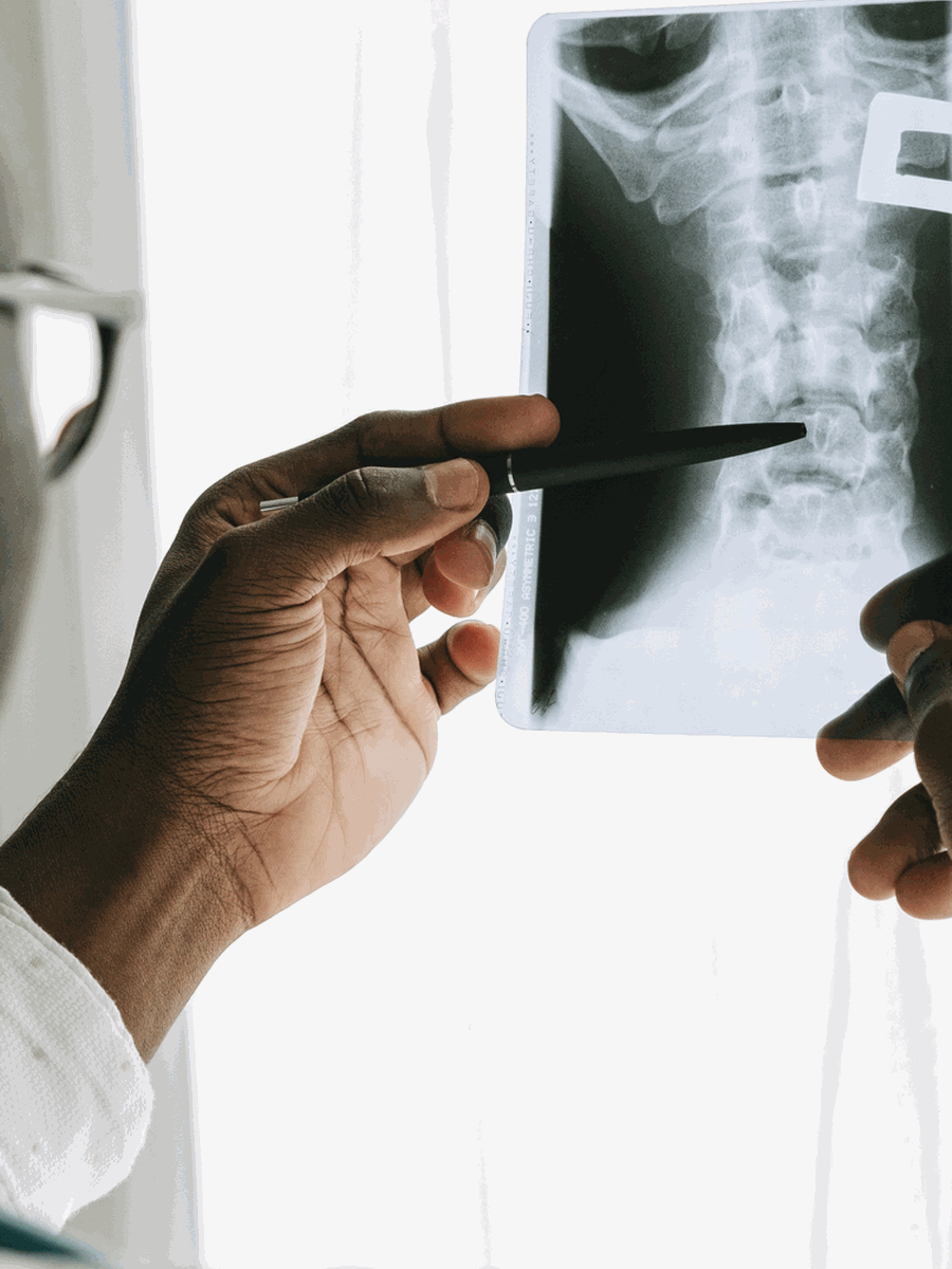
A person with new red-flag symptoms should attend an emergency department immediately rather than wait for an outpatient appointment. Assessment includes neurological examination, perineal sensation, bladder history, leg strength and urgent MRI. Post-void residual bladder measurement may add information but cannot safely rule out the syndrome alone. Digital rectal examination has limited sensitivity and should not delay imaging when suspicion is present. The ACR considers lumbar MRI without or without and with contrast usually appropriate for suspected cauda equina syndrome.
| Clinical pattern | Possible next step | Why it matters |
|---|---|---|
| New difficulty starting or sensing urination | Emergency assessment and urgent MRI | Incomplete syndrome may precede painless retention. |
| New saddle numbness or altered wiping sensation | Emergency assessment | Perineal sensory change is a core red flag. |
| Bilateral severe leg symptoms or progressive weakness | Urgent neurological evaluation | Multiple roots may be compressed. |
| MRI confirms compressive cauda equina syndrome | Emergency spinal-surgical pathway | Decompression is usually performed urgently. |

Treatment and decision-making
Management is not conservative when imaging confirms acute compressive cauda equina syndrome. The spinal team determines the urgency and type of decompression according to the cause, neurological stage and medical condition. Before confirmation, treatment focuses on rapid investigation, bladder monitoring, pain control and safe transfer. Steroids, manipulation, traction, exercise or repeated outpatient treatment should not delay MRI and surgical review. After decompression, recovery may require urology, bowel management, sexual-health care, rehabilitation, mobility support and psychological care.
How to interpret the finding in practice
The safest public message is simple: new bladder, bowel, sexual or saddle sensory change with back or leg symptoms should be treated as an emergency until assessed. A person should not try to decide at home whether the symptoms are severe enough.
Do not wait for complete retention
Earlier incomplete symptoms can still be serious.
Describe the change precisely
Explain what is new compared with normal bladder, bowel and sexual function.
Mention wiping sensation
Altered feeling when using toilet paper can be clinically important.
Report both legs
Bilateral pain, numbness or weakness increases concern.
Bring medication and surgical history
Anticoagulants, cancer, infection and recent procedures change the differential.
Go to emergency care
A clinic appointment or routine imaging request is not an adequate pathway.
How to monitor progress and avoid treatment errors
A credible treatment plan should define what improvement means before care begins. Pain intensity alone is not enough, because pain may fluctuate while neurological function, walking, sleep or daily activity changes in another direction. The diagnosis should also be reconsidered when the expected response does not occur.
Establish a baseline
Record symptom distribution, strength, sensation, walking, sleep, medication use and the activities that matter most.
Use a planned review point
Decide when progress will be evaluated rather than continuing automatically until a package is completed.
Separate relief from diagnosis
Temporary improvement after an injection, manual treatment or device does not prove that the proposed structure was the true cause.
Watch the neurological trajectory
New weakness, spreading numbness or balance change matters even if pain has decreased.
Revisit competing diagnoses
Hip, sacroiliac, vascular, inflammatory and postoperative causes may overlap with the initial explanation.
Escalate proportionately
Imaging, specialist referral or surgery should be considered when the risk or functional loss justifies it.
Common mistakes to avoid
- Treating the largest imaging abnormality rather than the clinically relevant lesion.
- Assuming that a diagnostic label remains correct when the symptom pattern changes.
- Repeating passive care despite no measurable functional progress.
- Using one negative test to exclude a condition that remains clinically plausible.
- Delaying urgent assessment to complete a predetermined treatment schedule.
- Generalizing results from a narrowly selected study population to every patient with a similar MRI term.
Shared decision-making is strongest when uncertainty is stated directly. A provider should explain what is confirmed, what remains probable, which alternatives still matter and what findings would change the plan. For complex or postoperative presentations, a second opinion from a clinician experienced with the specific condition may be more useful than adding another non-specific treatment.
Questions before choosing care
What is the leading diagnosis?
Ask which findings support it and which alternatives remain.
What test would change care?
Avoid testing that cannot alter the next step.
What result matters?
Track function, neurological safety and meaningful activity.
When should the plan change?
Define referral, stopping and escalation criteria.
Relevant specialized resources
Some resources may share ownership or editorial direction; this relationship is disclosed and the links are included for contextual relevance.
SOS Sciatique
Specialized warning-sign information about severe radicular symptoms.
Frequently asked questions
Is cauda equina syndrome common?
No. It is rare, but the consequences of missing it are serious.
Does urinary leakage always mean cauda equina syndrome?
No, but a new change with back or leg symptoms requires emergency assessment.
Can the syndrome begin with difficulty urinating rather than incontinence?
Yes.
What is saddle anaesthesia?
Reduced or altered sensation around the genitals, anus, buttocks and inner thighs.
Can sexual dysfunction be a warning sign?
Yes, when it is new and associated with spinal symptoms.
Does a normal post-void residual rule it out?
No single bladder measurement safely excludes all cases.
Is MRI required?
Urgent MRI is the usual investigation when the syndrome is suspected.
Can manipulation or decompression treatment be tried first?
No.
Does surgery guarantee full recovery?
No. Recovery depends on severity, duration, cause and individual factors.
What should I do if I have these symptoms now?
Go to the nearest emergency department immediately.
Sources consulted
- Hennessy et al. — International cauda equina guidelines review
- ACR Appropriateness Criteria — Low Back Pain, suspected cauda equina
- Somerset NHS — Cauda Equina Syndrome emergency pathway
- Royal Free London — Cauda Equina Syndrome patient guidance
Last editorial review: July 2026. General educational information only.
Dr. Sylvain Desforges, B.Sc., D.O., N.D., osteopath — The Spine Page
Continue exploring spine health
Use the condition and treatment hubs to compare related guides.
The Spine Page — The best treatments for your spinal problems — www.thespinepage.com
Topic index: Spinal emergencies and spinal-cord involvement
Explore this topic cluster by search intent. Each link leads to a distinct guide directly related to the subject.