





What Should You Do After a Spinal Decompression Session?
After a session, the goal is generally neither complete immobilization nor an immediate return to intense effort. Light activity, symptom monitoring and following the individualized plan are usually more coherent.
Instructions vary with the diagnosis, treated region, session response and usual activities. A short walk or previously well-tolerated movement may be encouraged, while heavy lifting, repeated twisting or unfamiliar training may be delayed when the area remains irritable.
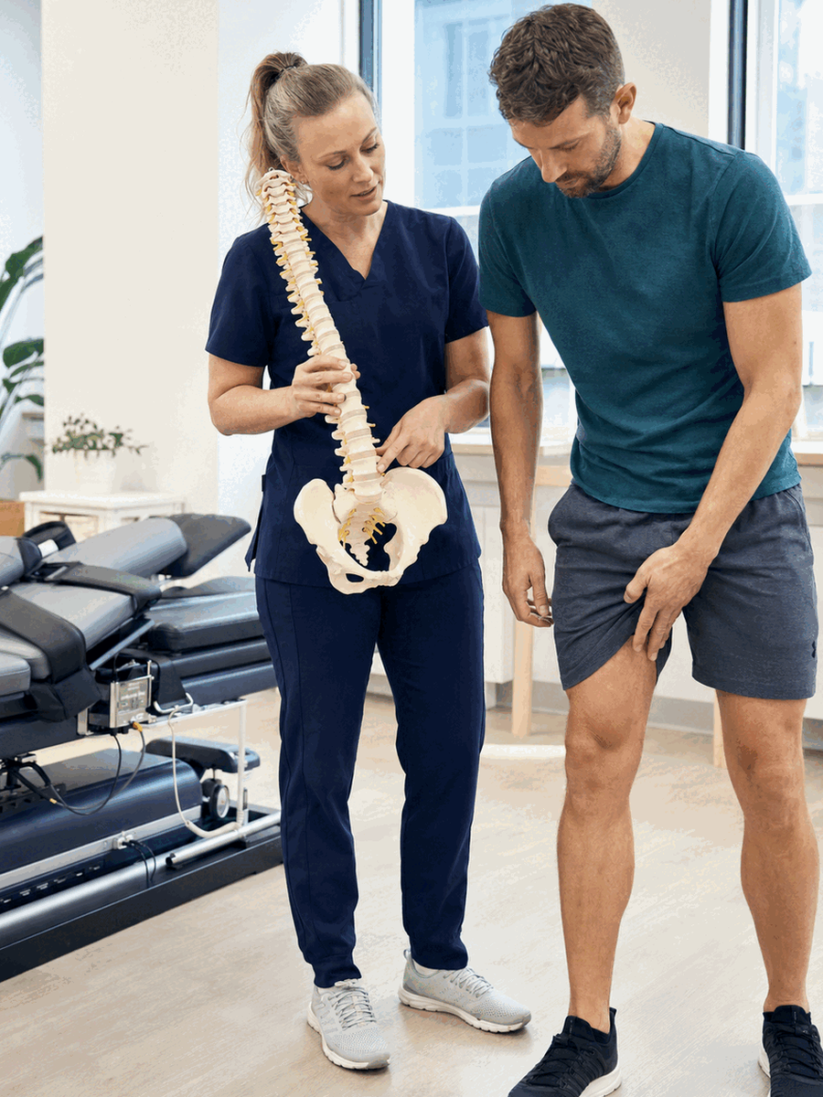
What Should You Do After a Spinal Decompression Session?
Instructions vary with the diagnosis, treated region, session response and usual activities. A short walk or previously well-tolerated movement may be encouraged, while heavy lifting, repeated twisting or unfamiliar training may be delayed when the area remains irritable.
It is useful to record symptom distribution rather than intensity alone. Pain centralizing toward the spine may mean something different from pain travelling farther into a limb. Any new weakness or sensory loss should be reported.
Clinical profile
Connect onset, territory, aggravating factors and relieving factors.
Neurology
Document strength, sensation, reflexes, balance, gait or dexterity as relevant.
Function
Measure sleep, work, driving, walking and priority activities.
Reassessment
Define when to continue, modify, stop or refer.
Clinical, functional and neurologic assessment
History covers onset, duration, distribution, positions, activities, prior care, medical background, medications and recent changes. Examination compares mobility, tolerance, strength, sensation, reflexes, gait, balance or dexterity as relevant.
Imaging is not automatic. It is most useful when it answers a question likely to change management, when neurologic deficit or a red flag is present, or before a targeted intervention. Imaging alone predicts neither pain nor treatment response.
| Clinical situation | Possible step | Why |
|---|---|---|
| Stable or improved response | Light activity as tolerated | Maintain mobility without provoking a major flare. |
| Mild soreness | Temporarily reduce load | Observe the duration and direction of the response. |
| Neurologic worsening | Communicate promptly | The plan and diagnosis should be reviewed. |
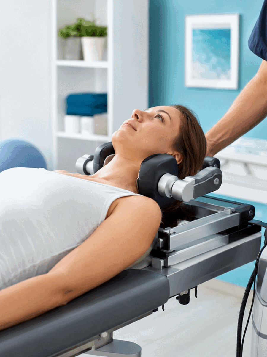

Build a measurable and reviewable trial
Before starting, select concrete measures: pain distribution, sleep, walking, sitting, driving, work, strength, sensation and an important activity. These markers help distinguish a temporary fluctuation from useful progress.
Continuation depends on a sufficiently durable favourable trend. Worsening, more distal symptoms, new weakness, persistent intolerance or lack of progress at the planned review point should lead to modifying, stopping or replacing the strategy.
It is useful to record symptom distribution rather than intensity alone. Pain centralizing toward the spine may mean something different from pain travelling farther into a limb. Any new weakness or sensory loss should be reported.
What to document before and during care
A useful plan turns observations into clinical questions and verifiable goals.
- Onset, duration, mechanism and evolution of symptoms.
- Territory, side, aggravating and relieving factors.
- Strength, sensation, reflexes, gait, balance or dexterity as relevant.
- Concordance among symptoms, function, examination and imaging when available.
- Functional goals and reassessment date.
- Stopping, escalation and urgent referral signs.
Track function rather than one isolated number
Follow-up combines pain intensity and distribution, strength, sensation, sleep, effort tolerance and participation. One good or bad day does not confirm an anatomic change.
Temporary relief may be useful without proving the proposed mechanism. Conversely, weakness, a fall, loss of dexterity or spreading numbness remains important even when pain fluctuates.
What is the working diagnosis?
Ask what supports it and what other causes remain possible.
What result will be measured?
Function and neurologic status complement a pain scale.
When will the plan be reviewed?
Set a date and criteria to continue, modify or stop.
What are the referral thresholds?
Clarify changes requiring imaging, medical review or emergency care.
Common mistakes to avoid
- Treating an image without checking clinical concordance.
- Promising universal repair or results.
- Continuing a protocol without useful measures.
- Ignoring differential diagnoses or warning signs.
- Confusing temporary relief with durable recovery.
- Delaying urgent referral to finish a protocol.
Questions to ask before starting
What is the working diagnosis?
Ask what supports it and what other causes remain possible.
What result will be measured?
Function and neurologic status complement a pain scale.
When will the plan be reviewed?
Set a date and criteria to continue, modify or stop.
What are the referral thresholds?
Clarify changes requiring imaging, medical review or emergency care.
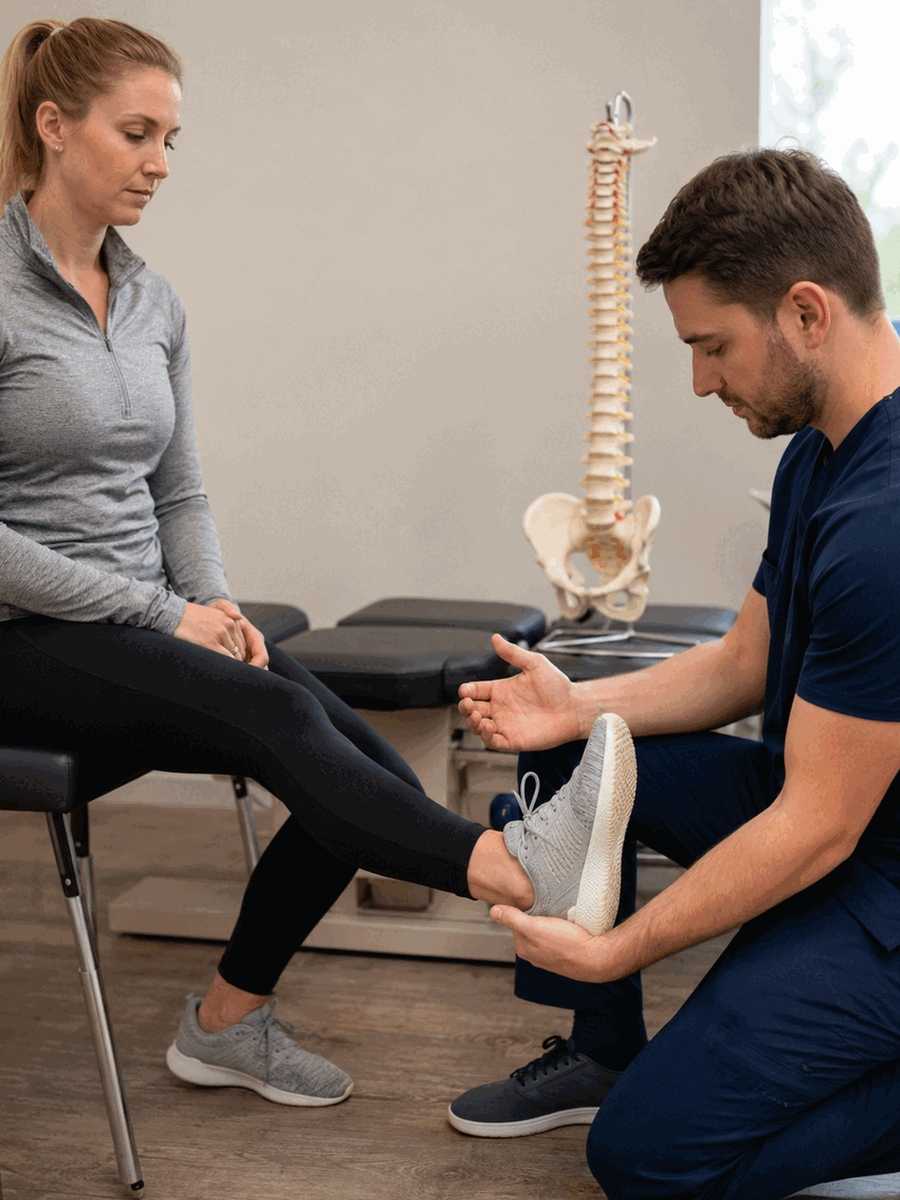
Related spinal decompression guides
When Can Improvement Begin With Spinal Decompression?
Explore this complementary search intent within the thematic silo.
No Improvement With Spinal Decompression: When to Reassess or Change Strategy
Explore this complementary search intent within the thematic silo.
Spinal Decompression, Exercise and Rehabilitation: How to Integrate Them
Explore this complementary search intent within the thematic silo.
Disclosed clinical resource
Readers who wish to verify a clinical service may consult this separate disclosed resource. This link does not replace comparing options or urgent referral.
Clinique TAGMED
Information on assessment and selected nonsurgical care that may be discussed for certain disc-related or radicular conditions.
Frequently asked questions
Is spinal decompression recommended for everyone?
No. It may be discussed for selected profiles after triage and examination. General guidance for low back and neck pain does not support traction as a universal solution.
Is MRI required?
No. Imaging is most useful when it can change management, when neurologic deficit or red flags are present, or before a targeted procedure.
How should meaningful improvement be measured?
Combine pain intensity and distribution with strength, sensation, sleep, walking, positional tolerance and participation in important activities.
Is lower pain immediately after a session enough?
No. Brief relief is of interest, but it should become sufficiently durable and be accompanied by functional or neurologic improvement.
What if symptoms travel farther down the limb?
Report this promptly. More distal pain or numbness may indicate increased irritation and justify changing the plan.
Can decompression be combined with exercise?
Yes, when exercises are selected according to diagnosis and tolerance. Active progression is generally important for rebuilding capacity.
How many sessions are needed?
There is no universal number. A credible trial defines a time frame, baseline measures, stopping criteria and a reassessment date.
When should treatment be stopped?
With repeated worsening, new weakness, persistent intolerance, a red flag or insufficient progress at the planned review point.
Does decompression permanently repair a disc?
Evidence does not support promising permanent anatomic repair. The clinical goal is measurable improvement in symptoms and function.
When is urgent assessment needed?
Rapidly progressive weakness, saddle numbness, bladder or bowel changes, fever with spinal pain, major trauma or myelopathic signs require prompt assessment.
Principal clinical sources
- NICE — Low back pain and sciatica in over 16s
- Cochrane — Traction for low-back pain
- World Health Organization — non-surgical management of chronic primary low back pain
- American College of Radiology — Low Back Pain
Last editorial review: August 2026. General educational information only.
Dr. Sylvain Desforges, B.Sc., D.O., N.D., osteopath – The Spine Page
Continue your research on spinal decompression
Compare the decision pages, general guide and spine conditions to maintain a coherent and prudent overview.
Dr. Sylvain Desforges, B.Sc., D.O., N.D., osteopath