





Spinal infection, discitis and spondylodiscitis: warning signs and diagnosis
Spinal infection may involve the vertebral body, disc space, epidural space or surrounding soft tissues. It can resemble degenerative back pain and may occur without fever, so persistent severe pain with infection risk factors deserves prompt assessment.
Native vertebral osteomyelitis commonly results from bacteria spreading through the bloodstream to adjacent vertebral endplates and the disc. Staphylococcus aureus is a common organism, but tuberculosis, Brucella, fungi and other bacteria occur in specific epidemiological and immune contexts. Infection may follow bloodstream infection, endocarditis, urinary or skin infection, intravenous drug use, spinal surgery, injection or an implanted device.
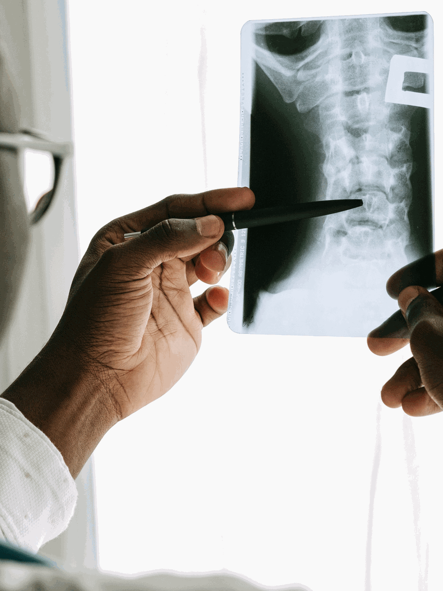
Assessment and differential diagnosis
IDSA recommends medical and neurological examination, two sets of blood cultures, ESR and CRP, and spine MRI when native vertebral osteomyelitis is suspected. MRI without and with contrast is usually appropriate according to the ACR. If blood cultures do not identify an organism, image-guided biopsy of the disc or endplate may be required before antibiotics, provided the patient is stable. Empiric antimicrobial therapy should not be delayed in sepsis, haemodynamic instability or neurological compromise.
| Clinical pattern | Possible interpretation | Why it matters |
|---|---|---|
| New worsening back pain with fever or elevated ESR/CRP | Suspect vertebral infection | Obtain cultures and MRI. |
| New spinal pain after Staphylococcus aureus bloodstream infection | High clinical concern | Prompt infectious-disease assessment is appropriate. |
| Neurological loss or epidural abscess | Emergency surgical and antimicrobial pathway | Spinal cord or cauda equina function may be threatened. |
| Favourable response to treatment | Clinical and laboratory monitoring | Routine follow-up MRI is not always recommended. |
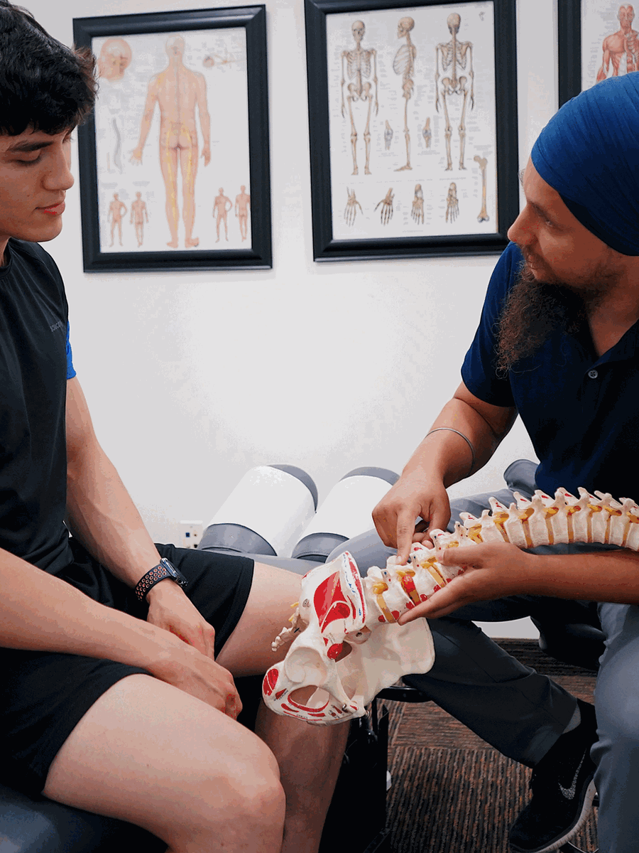

Treatment and decision-making
Treatment is directed by the identified organism and usually involves prolonged antimicrobial therapy. Drainage, debridement or stabilization may be required for neurological compromise, large abscess, progressive deformity, instability, uncontrolled sepsis or treatment failure. Rehabilitation and mobilization are introduced according to stability and medical response. Massage, manipulation, traction or decompression technology is inappropriate over an untreated spinal infection.
How to interpret progress and avoid common mistakes
A credible plan should explain not only what treatment may be tried, but also how the diagnosis will be reconsidered if the expected response does not occur. Pain intensity can fluctuate for reasons that do not necessarily reflect tissue damage, while neurological or systemic deterioration can occur even when pain changes little. For that reason, progress should be judged with several measures rather than one daily pain score.
Establish a baseline
Record symptom distribution, aggravating activities, walking or sitting tolerance, sleep, medication use and the neurological findings relevant to this condition.
Choose a meaningful goal
Define a practical target such as walking farther, sleeping through the night, returning to work, tolerating sitting or recovering strength.
Use a planned review point
Decide in advance when the response will be reviewed rather than continuing indefinitely because a treatment package has not been completed.
Separate symptom relief from diagnosis
A temporary improvement after medication, manual care, injection or a device does not by itself prove that the proposed structure was the true pain source.
Watch the overall trajectory
Small day-to-day fluctuations are common. The more important question is whether function, neurological safety and participation are improving over time.
Escalate when the pattern changes
New weakness, systemic illness, vascular change, severe night pain or loss of bladder or bowel control requires a different pathway rather than more of the same treatment.
Assumptions that should be avoided
- Do not assume that every abnormal image is symptomatic.
- Do not assume that one negative test excludes the condition when clinical suspicion remains.
- Do not use treatment response as the only diagnostic test.
- Do not delay referral simply to finish a predetermined number of visits.
- Do not generalize results from a narrowly selected trial population to every person with a similar label.
- Do not interpret the absence of severe pain as proof that neurological or systemic risk is absent.
Shared decision-making is strongest when uncertainty is stated directly. A provider should be able to explain which findings support the working diagnosis, which competing diagnoses remain possible, what evidence applies to the proposed intervention, and what would trigger imaging, laboratory testing, specialist referral or a change of plan. When several conditions can produce similar symptoms, the safest approach is often staged: first exclude urgent disease, then identify the most likely mechanism, begin proportionate care and reassess against objective goals.
Questions before choosing care
What is the leading diagnosis?
Ask which findings support it and which alternatives remain.
What test would change care?
Avoid testing that cannot alter the next step.
What result matters?
Track function, neurological safety and meaningful activity.
When should the plan change?
Define referral, stopping and escalation criteria.

Relevant specialized resources
Some resources may share ownership or editorial direction; this relationship is disclosed and the links are included for contextual relevance.
Dr Sylvain Desforges
Professional information about warning signs and appropriate referral.
Frequently asked questions
Can spinal infection occur without fever?
Yes.
Is discitis the same as vertebral osteomyelitis?
They often coexist and the term spondylodiscitis describes combined disc and vertebral infection.
Which blood tests are used?
Blood cultures, ESR and CRP are commonly obtained.
Which imaging test is preferred?
MRI is generally preferred.
Why is biopsy sometimes needed?
To identify the organism when blood cultures are not diagnostic.
Should antibiotics start before biopsy?
Not always in a stable patient, but they should not be delayed in sepsis or neurological compromise.
How long is treatment?
Duration depends on the organism, response and complications and is directed by specialists.
Is follow-up MRI always needed?
No, not when clinical and laboratory response is favourable.
Can manipulation or traction be used?
Not over an untreated or unstable spinal infection.
Does The Spine Page diagnose infection?
No.
Sources consulted
- IDSA — Native Vertebral Osteomyelitis Guideline
- ACR Appropriateness Criteria — Suspected Spine Infection
Last editorial review: July 2026. General educational information only.
Dr. Sylvain Desforges, B.Sc., D.O., N.D., osteopath — The Spine Page
Continue exploring spine health
Use the condition and treatment hubs to compare related guides.
The Spine Page — The best treatments for your spinal problems — www.thespinepage.com