





L4-L5 and L5-S1 Degenerative Disc Disease: Levels, Roots and Symptoms
L4-L5 and L5-S1 commonly show degenerative changes. The disc level does not directly match the name of the irritated nerve root, which explains many misunderstandings in reports and patient searches.
An L4-L5 problem may often involve the L5 root, whereas an L5-S1 problem may involve S1. Actual distribution, strength and reflexes remain essential.
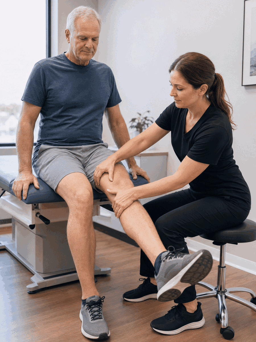
L4-L5 and L5-S1 Degenerative Disc Disease: Levels, Roots and Symptoms
Pain toward the great toe, difficulty lifting the foot, pain along the outer foot or difficulty rising onto the toes may guide the examination without establishing the diagnosis by themselves.
The goal is to turn this topic into testable clinical questions, baseline measures and explicit reassessment criteria rather than a universal promise.
Clinical profile
Relate onset, distribution and modifying factors.
Neurology
Document strength, sensation, reflexes and function.
Participation
Measure sleep, work, walking and important activities.
Reassessment
Plan when to continue, modify or refer.
Clinical, functional and neurologic assessment
History clarifies onset, duration, distribution, provoking activities, prior care, recent changes and relevant medical history. Examination compares mobility, function, strength, sensation, reflexes and differential diagnoses according to the region.
Imaging or additional tests are not automatic. They become more useful when they answer a question likely to change management, when a deficit or red flag is present, or before a targeted procedure.
| Clinical situation | Possible step | Why |
|---|---|---|
| Stable symptoms without progressive deficit | Graded plan and baseline measure | Preserve function and avoid overmedicalization. |
| Persistent or atypical symptoms | Targeted diagnostic review | Check another cause or a specific indication. |
| Weakness or red flag | Proportionate referral and testing | Priority may change quickly. |


Building a measurable and revisable plan
The baseline should include an activity that matters to the person, symptom distribution, tolerance to positions or effort, sleep and relevant neurologic findings.
The plan should be based on the likely involved root, function and deficit stability rather than only the level named in the report.
Each intervention should have a goal, trial duration, stopping criteria and reassessment date. Continued care should be justified by sufficiently durable and functional improvement.
What to document before and during care
A useful plan converts observations into clinical questions and verifiable goals.
- Onset, duration, mechanism and symptom evolution.
- Distribution, side, provoking factors and relief.
- Strength, sensation, reflexes, walking, balance or dexterity by region.
- Concordance among symptoms, function, examination and imaging when available.
- Functional goal and reassessment date.
- Stopping, escalation and urgent referral signs.
Following function rather than one isolated number
Follow-up combines pain, distribution, strength, sensation, sleep, tolerance and participation. A one-day fluctuation does not by itself prove an anatomic change.
Temporary relief does not necessarily confirm the proposed mechanism. Conversely, weakness, a fall, loss of dexterity or spreading numbness remains important even when pain decreases.
What is the working diagnosis?
Ask what supports it and which other causes remain possible.
What outcome will be measured?
Function and neurology complement a pain scale.
When will the plan be reviewed?
Set a time and criteria to continue, modify or stop.
What are the referral thresholds?
Clarify changes requiring imaging, medical review or emergency care.
Common errors to avoid
- Treating an imaging term without checking concordance.
- Promising a universal anatomic or clinical result.
- Repeating an intervention without a functional measure.
- Ignoring important differential diagnoses.
- Confusing temporary relief with neurologic recovery.
- Delaying urgent referral to finish a protocol.
Questions to ask before starting
What is the working diagnosis?
Ask what supports it and which other causes remain possible.
What outcome will be measured?
Function and neurology complement a pain scale.
When will the plan be reviewed?
Set a time and criteria to continue, modify or stop.
What are the referral thresholds?
Clarify changes requiring imaging, medical review or emergency care.
Related guides in the degenerative disc disease silo
C5-C6 and C6-C7 Degenerative Disc Disease: Neck, Arm and Hand Symptoms
Explore this complementary intent within the topic silo.
Degenerative Disc Disease or Facet Arthritis: Distinguishing Possible Sources
Explore this complementary intent within the topic silo.
Exercise and Activity with Degenerative Disc Disease: Symptom-Guided Progression
Explore this complementary intent within the topic silo.
Disclosed clinical resource
Readers who wish to verify a clinical service may consult this separate, disclosed resource. The link does not replace comparison of options or urgent referral when required.
TAGMED Clinic
Information about assessment and selected non-surgical care that may be discussed for selected disc-related problems.
Frequently asked questions
What exactly does this term mean?
It describes an observation or hypothesis that must be related to symptoms, function and examination. By itself, it is not a treatment indication.
Does imaging always explain symptoms?
No. Many degenerative changes are common in people without pain. Clinical concordance remains essential.
Is MRI always required?
No. It is most useful when it can change management, when deficits or red flags are present, or before a targeted procedure.
Can a person remain active?
Often yes, with adaptation and progression. Neurologic or functional worsening requires reassessment.
Is spinal decompression suitable for everyone?
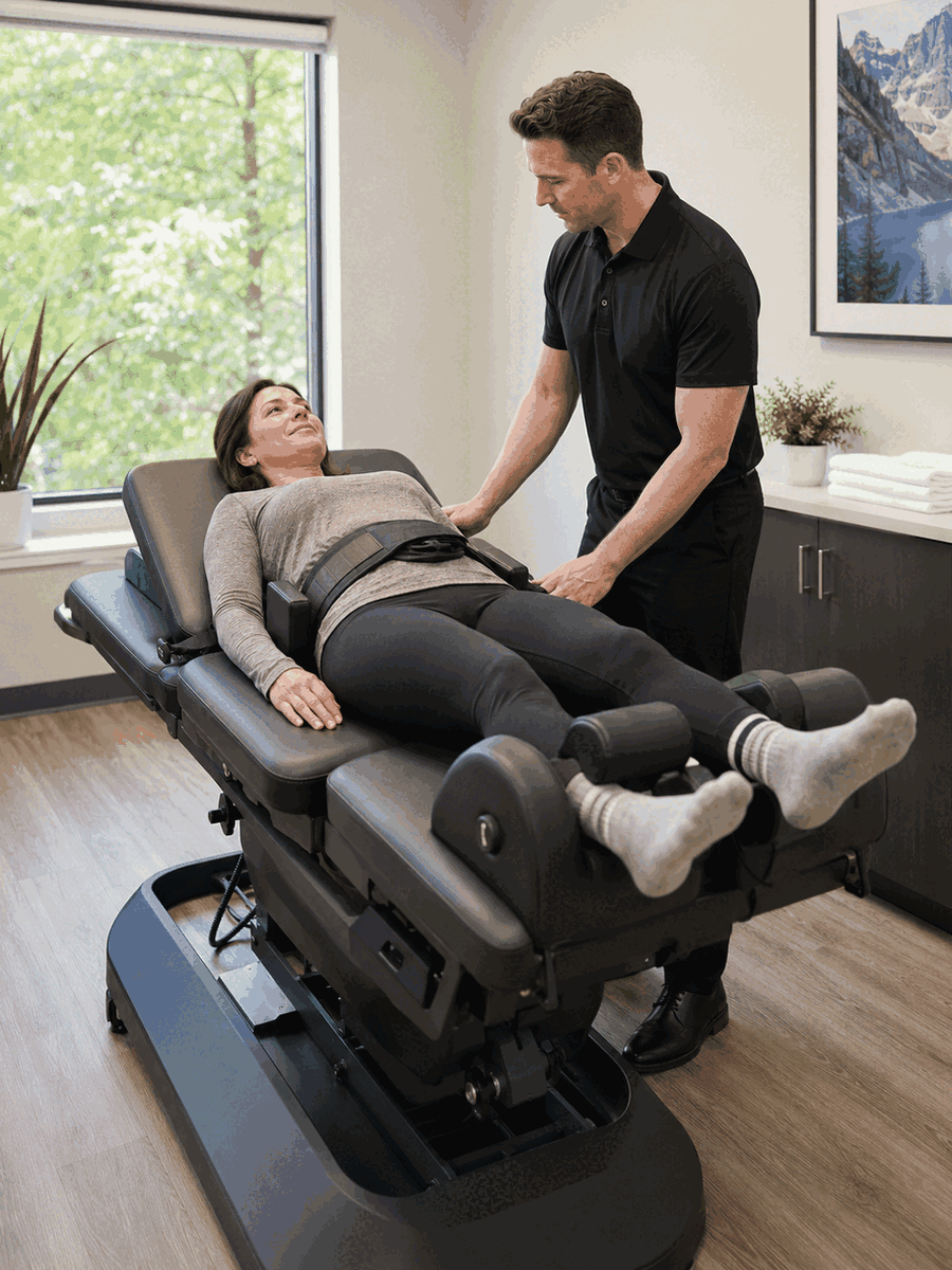
No. It applies to selected mechanical or compressive profiles and does not replace triage or an active plan.
How should progress be measured?
Combine a functional activity, symptom distribution, sleep, tolerance and relevant neurologic findings.
How long should a treatment be tried?
It depends on the diagnosis and intervention. A reassessment date and modification criteria should be set in advance.
Is pain improvement enough?
No. Strength, sensation, walking, dexterity, sleep and participation may change differently.
Is surgery automatically required?
No. Many stable presentations first receive conservative care. An emergency, progressive deficit or specific indication may justify surgical review.
When is urgent assessment needed?
Foot drop, progressive weakness, saddle anesthesia, bladder or bowel changes or bilateral symptoms requires urgent assessment.
Main clinical sources
- NICE — Low back pain and sciatica in over 16s
- WHO — Guideline for non-surgical management of chronic primary low back pain
- American College of Radiology — Low Back Pain
Last editorial review: August 2026. General educational information only.
Dr. Sylvain Desforges, B.Sc., D.O., N.D., osteopath – The Spine Page
Continue your research on degenerative disc disease
Compare related guides and return to the general page to maintain a coherent overview.
Dr. Sylvain Desforges, B.Sc., D.O., N.D., osteopath