





Herniated disc assessment in Terrebonne: examination and imaging
A credible assessment does not merely look for a herniation: it tests whether the disc finding truly explains symptoms and whether a deficit changes urgency.
History clarifies onset, mechanism, distribution, aggravating positions, previous care and changes in strength or sensation.
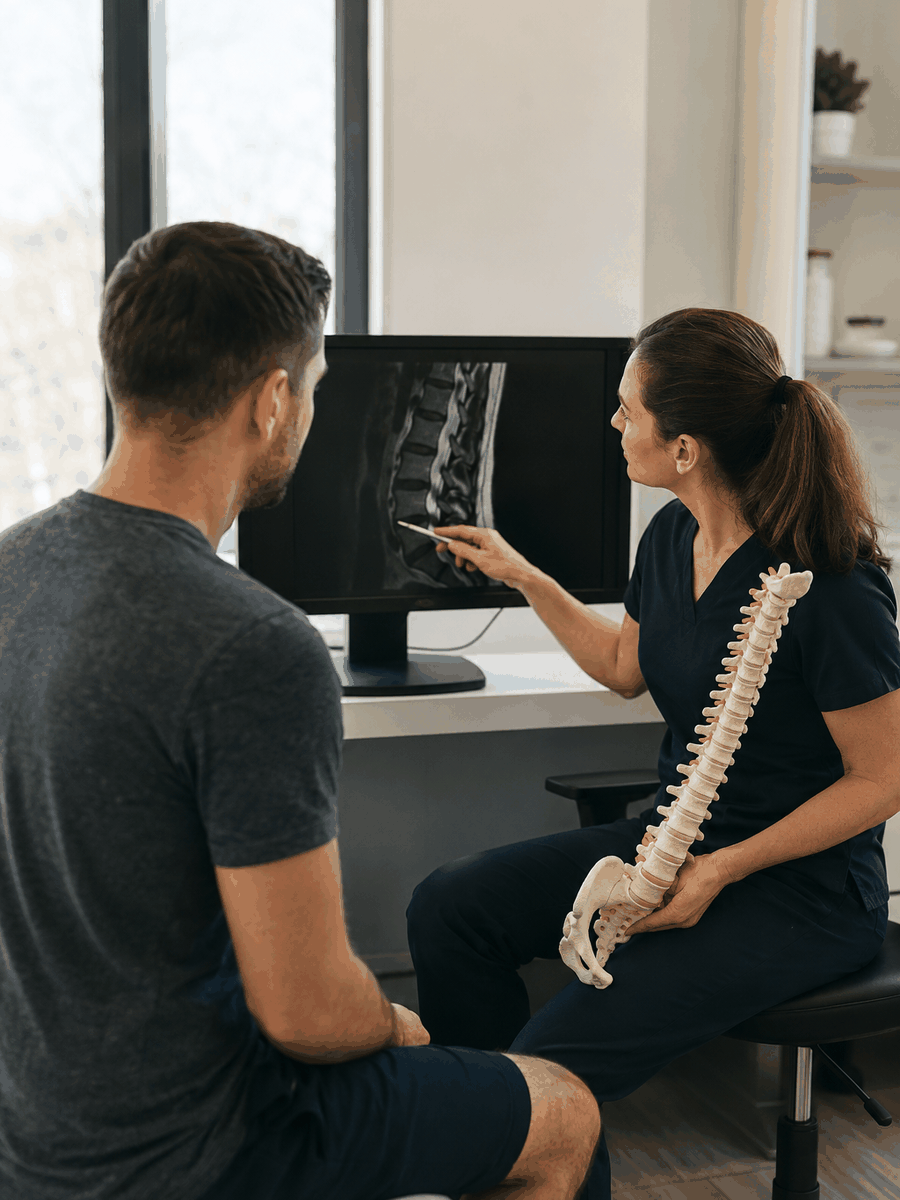
Clinical assessment and decision points
Assessment documents onset, distribution, modifying positions, previous care and changes in strength or sensation. It also checks for conditions that can mimic a symptomatic herniation.
Imaging is not automatic. It becomes more relevant with warning signs, before a contemplated procedure, or when persistent symptoms make it likely that the result will change management.
| Clinical situation | Possible next step | Why it matters |
|---|---|---|
| Stable symptoms without deficit | Graduated care and follow-up | Support function without overmedicalizing. |
| Numbness or weakness | Focused neurological examination | Progression changes urgency and referral. |
| Persistent or atypical symptoms | Diagnostic review and selective imaging | Another cause or targeted intervention may need discussion. |


When does imaging change management?
MRI is not automatically required for a first stable episode. It becomes more relevant with deficit, warning signs, persistent symptoms or before a targeted intervention.
The report should be compared with the images when available and, most importantly, with the clinical presentation. A protrusion on the wrong side or at a non-corresponding level should not dictate treatment.
Elements to document in a credible plan
A useful plan turns a diagnosis into observable goals. It specifies what should improve, what will be monitored and what will require a different pathway.
- Record the side and exact symptom distribution.
- Compare strength and sensation with the opposite side.
- Identify positions and loads that aggravate or centralize symptoms.
- Measure one meaningful, repeatable activity.
- Set a review date before beginning.
- Define stopping signs and referral criteria.
Track progress and avoid automatic continuation
Pain intensity is one measure among others. Distribution, strength, sensation, gait, sleep, positional tolerance and participation provide a more reliable picture.
Temporary relief does not necessarily confirm the proposed mechanism. Conversely, one difficult day does not prove deterioration. Trend, function and neurological status should be interpreted together.
Establish a baseline
Document distribution, strength, sensation, tolerance and one important activity.
Schedule reassessment
Decide in advance when outcomes will be compared to avoid automatic continuation.
Monitor neurology
New or progressive weakness matters even when pain decreases.
Compare pain and function
Relief does not prove functional or neurological recovery.
Review other causes
The hip, a joint, a peripheral nerve or systemic condition can imitate the presentation.
Escalate proportionately
Imaging, injection or surgery are discussed when risk or functional loss justifies them.
Common errors to avoid
- Treating a word from the MRI report without checking side and distribution.
- Promising a fixed visit count before establishing a baseline.
- Confusing short-term relief with neurological recovery.
- Continuing passive care despite no functional progress.
- Ignoring weakness because pain fluctuates.
- Delaying urgent referral to finish a protocol.
Questions to ask before starting
What is the working diagnosis?
Ask which findings support it and which alternative causes remain possible.
Which outcome will be measured?
Walking, sleep, strength and one meaningful activity complement the pain score.
When will the plan be reviewed?
Set a time and criteria to continue, change or stop.
What are the referral thresholds?
Clarify which changes require imaging, medical review or urgent care.

Related guides in the Terrebonne cluster
Herniated disc treatment in Terrebonne: assessment and options
Explore this complementary intent within the Terrebonne cluster.
Non-surgical herniated disc treatment in Terrebonne
Explore this complementary intent within the Terrebonne cluster.
Spinal decompression for a herniated disc in Terrebonne
Explore this complementary intent within the Terrebonne cluster.
Specialized local resource
Readers who wish to verify a local clinical offering can consult the separate, disclosed resource below. This link does not replace comparison of options.
Clinique TAGMED – Terrebonne
Information about assessment and non-surgical care offered in Terrebonne for selected disc herniations.
Frequently asked questions
What is the first goal when considering herniated disc assessment in terrebonne: examination and imaging?
The first goal is to establish concordance between symptoms, examination and the working diagnosis, then measure function and neurological status.
How long should an option be tried before review?
Timing depends on diagnosis and intervention. A credible plan sets a review date and modification criteria rather than one universal visit count.
Is MRI required before every treatment?
No. It is most useful when it can change management, when a deficit or warning sign is present, or before a targeted intervention.
Is decompression suitable for every herniation?
No. Selection depends on profile, spinal region, tolerance, risks and contraindications. It does not replace neurological triage.
How can two Terrebonne treatment plans be compared?
Compare the diagnostic hypothesis, functional goals, evidence explained, risks, review schedule and referral criteria.
Can a person remain active with a herniated disc?
Often yes, with modification and progression. Activity should be reduced or reassessed if symptoms travel farther or strength deteriorates.
Does a disc herniation on MRI always explain the pain?
No. A disc abnormality should match the affected side, symptom distribution, neurological examination and clinical course. Some herniations are incidental.
Is surgery automatically required?
No. Many stable presentations are initially managed conservatively. A neurological emergency, progressive deficit or well-documented failure may nevertheless justify surgical assessment.
Does bed rest speed recovery?
Prolonged bed rest is generally discouraged. Modified, graduated and monitored activity is usually preferable when neurological status is stable.
When is urgent assessment required?
With new or rapidly progressive weakness, foot drop, saddle numbness, bladder or bowel change, fever or significant trauma.
Main clinical sources
- North American Spine Society – Lumbar Disc Herniation with Radiculopathy
- American College of Radiology – Low Back Pain Appropriateness Criteria
- NICE – Low back pain and sciatica in over 16s
Last editorial review: August 2026. General educational information only.
Dr. Sylvain Desforges, B.Sc., D.O., N.D., osteopath – The Spine Page
Continue researching disc herniation
Compare related guides and return to the pillar page for a complete view of herniated disc treatment in Terrebonne.
The Spine Page – The best treatments for your spinal problems – www.thespinepage.com