

A vertebral lamina is one of two bony plates at the back of the vertebral arch. Together, the laminae contribute to the posterior covering of the spinal canal and meet near the spinous process; their shape varies by spinal level.
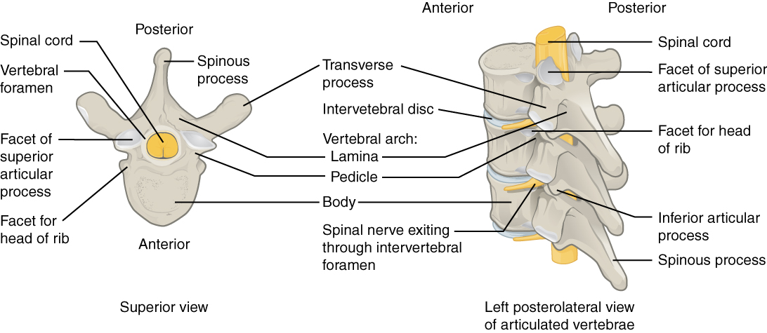
OpenStax College — CC BY 3.0. Image source.

Structure and function
The laminae sit behind the neural structures within the canal. They extend from the articular regions toward the midline. Ligamenta flava connect laminae of neighbouring vertebrae. This arrangement combines bony protection, ligament attachments and movement between spinal segments, rather than forming an immovable shell around the entire spine.
Possible manifestations
A lamina may fracture during trauma or have a developmental variation. It is also an important surgical landmark. Pain in the middle of the back cannot establish that the lamina is the source: muscles, joints and other tissues may produce discomfort in the same area. A scan finding needs to fit the clinical presentation.
Assessment and management
CT can assess bony continuity in detail. MRI evaluates canal contents and soft tissues when symptoms justify it. Laminectomy removes part or all of a lamina; laminotomy creates a more limited opening. These procedures address selected compression problems and are not a general treatment for every episode of spinal pain.
Role of TAGMED care
At TAGMED, a previous laminectomy requires understanding the procedure, recovery and surgeon’s restrictions. Physical care may subsequently be adapted to functional goals. It does not aim to regrow removed bone, and persistent postoperative symptoms deserve assessment before additional mechanical treatments.
Warning signs
Pain after trauma with weakness, walking changes or urinary dysfunction requires urgent assessment. Following surgery, new weakness or bladder or bowel dysfunction requires immediate medical attention rather than waiting for a scheduled physical treatment appointment.
Frequently asked questions
Is it a disc?
No. The lamina is bone behind the spinal canal.
Does the lamina contain the cord?
It forms part of the bony protection surrounding the canal.
Does removal always require fusion?
No. Stability and the extent of surgery determine that decision.
Is partial absence always traumatic?
No. It may be developmental or result from surgery.
Is MRI always necessary?
No. Imaging should answer a specific clinical question.
Conceptual anatomical and clinical relationships; not drawn to scale.
Medical references
- OpenStax — Rice University. The Vertebral Column.
- AAOS — OrthoInfo. Fractures of the Thoracic and Lumbar Spine.
- ACR — RSNA. Spine MRI.
References document the topic; their inclusion does not imply endorsement of TAGMED services by those organizations.
Educational information from The Spine Page. This entry does not replace an individual assessment. Documentary version: October 12, 2026. Sylvain Desforges, B.Sc., D.O., N.D., osteopath and naturopath.
TAGMED clinic contact details
For non-urgent consultations and information about available services.
Clinique TAGMED — Terrebonne
1150, rue Lévis, Suite 200
Terrebonne (Québec) J6W 5S6
450‑704‑4447
Clinique TAGMED — Montréal
1140, avenue Beaumont
Mont-Royal (Québec) H3P 3E5
1‑877‑672‑9060