

Bony outgrowth that can develop near a joint or vertebral endplate during degenerative change.
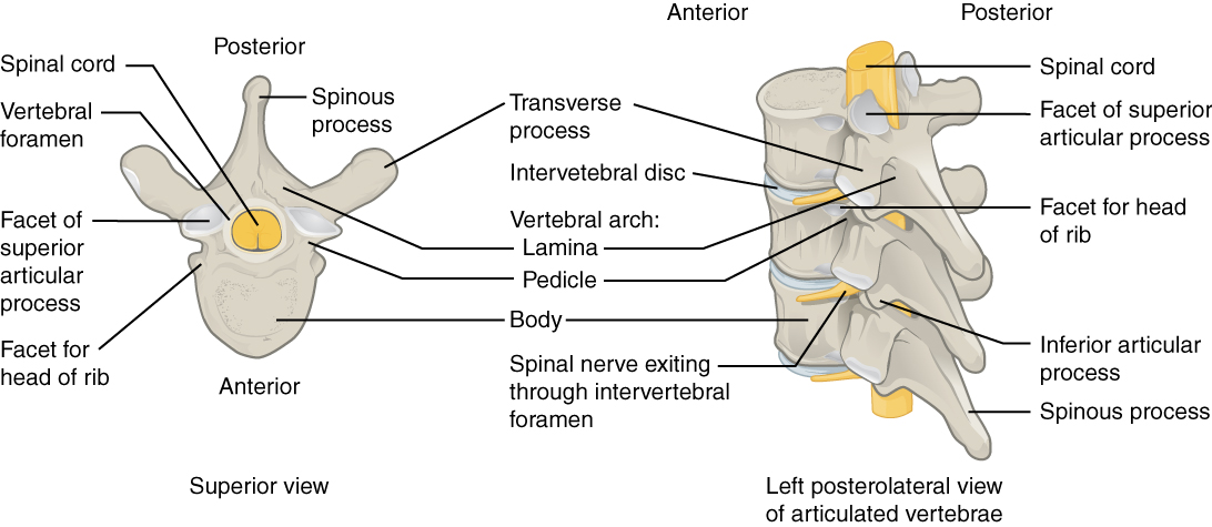
OpenStax College — CC BY 3.0. Image source.

Understanding the mechanism
An osteophyte, or bone spur, is a bony outgrowth near a joint or vertebral endplate, often associated with degeneration. It represents bone remodelling rather than a displaced piece of disc. Its presence does not automatically indicate pain or danger. Location, the effect on neighbouring tissues and the clinical context determine whether it has practical significance.
Symptoms and warning signs
Many osteophytes cause no symptoms. Some contribute to stiffness or narrowing of a foramen or spinal canal, potentially affecting a root or the spinal cord. Progressive weakness, altered walking or unusual swallowing difficulty warrants assessment, although other causes must also be considered. A visible spur cannot independently establish the explanation for any of these symptoms.
Diagnosis and investigations
Radiographs and CT show bone well. MRI provides more information about roots, the spinal cord and soft tissues when neurological symptoms are present. The clinician checks whether the spinal level and side match the presentation. An incidental finding does not necessarily require additional tests; comparison with earlier imaging can sometimes clarify whether a change is clinically relevant.
Treatment and outlook
Management targets any clinical consequences rather than the term osteophyte itself. Appropriate activity, strengthening and medical pain management may be sufficient for some presentations. Surgery is considered only for particular indications, such as significant symptomatic neural compression. Conservative treatment aims to improve function even if the spur remains visible. Image normalization is not a necessary measure of successful care.
The role of TAGMED care
At TAGMED, it is important to explain that traction does not dissolve bone and shockwave therapy is not intended to break spinal osteophytes. Laser and manual therapy are not established ways to remove them. Any treatment should address assessed associated symptoms, with medical referral when neurological compression or another complication is suspected rather than promising to eliminate the imaging finding.
Practical follow-up
Keeping the report and identifying the level involved supports a precise discussion of the spur's significance. Comparing scans is useful when it answers a clinical question, but it does not replace assessment of nerve function or the person's actual limitations.
Frequently asked questions
Is a bone spur a bone tumour?
A degenerative osteophyte is not synonymous with cancer; atypical findings still require their own assessment.
Does it necessarily explain my pain?
No. The relationship between the scan and symptoms must be checked.
Can treatment dissolve it?
Routine conservative care has not been shown to dissolve spinal osteophytes.
Does it need removal?
Only in selected specialist indications; many remain symptom-free.
Why monitor strength and walking?
The effect on a root or the cord matters more than size alone.
Conceptual diagram: the steps depend on clinical findings and do not provide an individual diagnosis.
Medical references
- NIAMS. Osteoarthritis.
- American Academy of Orthopaedic Surgeons. Cervical Radiculopathy (Pinched Nerve).
- American Academy of Orthopaedic Surgeons. Cervical Spondylotic Myelopathy.
References document the topic; their inclusion does not imply endorsement of TAGMED services by those organizations.
Educational information from The Spine Page. This entry does not replace an individual assessment. Documentary version: October 12, 2026. Sylvain Desforges, B.Sc., D.O., N.D., osteopath and naturopath.
TAGMED clinic contact details
For non-urgent consultations and information about available services.
Clinique TAGMED — Terrebonne
1150, rue Lévis, Suite 200
Terrebonne (Québec) J6W 5S6
450‑704‑4447
Clinique TAGMED — Montréal
1140, avenue Beaumont
Mont-Royal (Québec) H3P 3E5
1‑877‑672‑9060