

Imaging using magnetic fields and radio waves, particularly useful for discs, the spinal cord and soft tissues.
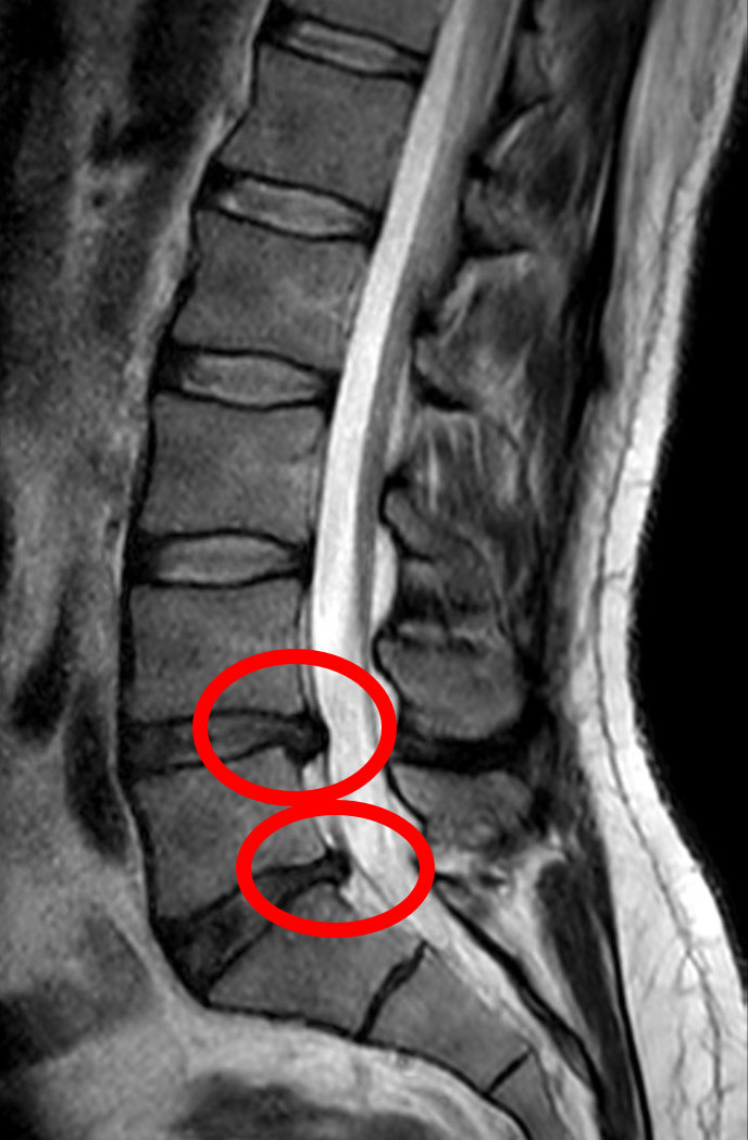
Miguel Tremblay (Dirac) — CC0 1.0. Image source.

Imaging discs, nerves and the cord without x-rays
MRI uses a magnetic field and radio waves. It provides detailed information about discs, the spinal cord, nerve roots and many soft tissues. It may be indicated for neurological deficits, suspected infection or tumour, or planning an intervention. It is not routinely necessary for an uncomplicated episode of back pain.
The person lies in a noisy scanner and needs to remain relatively still. Some examinations use contrast material, but many do not. Before any MRI, report implants, medical devices, metallic foreign bodies and possible pregnancy. Also report kidney disease, particularly if contrast material is being considered. Claustrophobia can be discussed beforehand. A radiologist interprets the images, and the responsible clinician assesses their clinical relevance. An implanted device is not automatically a prohibition, but its exact safety conditions must be checked by the imaging team.
Indications and interpretation
Before ordering the test, the clinician should identify the question it needs to answer and how the result could change decisions. Routine imaging for low back pain without warning signs does not necessarily improve care. A result describes structures; it does not measure all aspects of pain or a person's capacity to recover. Age, previous conditions, the affected side and the physical examination help distinguish a relevant abnormality from an incidental finding. More visible detail is useful only when interpreted appropriately.
After the results
The responsible clinician should explain which findings need action, follow-up or no particular intervention. Bringing previous studies can prevent unnecessary repetition. At TAGMED, imaging can help select or exclude particular care options; it cannot establish that traction, laser treatment or manual care will work. Decisions remain clinical and must consider contraindications. Progressive weakness, altered saddle sensation or a new inability to urinate must not wait for a routine imaging appointment: these symptoms need emergency assessment. Follow the preparation and safety instructions supplied by the imaging centre. Ask how and when results will be communicated rather than assuming that silence means everything was normal.
Frequently asked questions
Does MRI use x-rays?
No. It uses a magnetic field and radio waves, with specific safety precautions.
Does an abnormality necessarily explain pain?
No. Agreement with symptoms and examination findings must be checked.
Should previous results be brought?
Yes when available; comparison may prevent unnecessary repeat studies.
Who explains the report?
The responsible clinician interprets it within the whole clinical picture.
Does the test determine treatment?
It contributes to decisions but does not replace clinical assessment and discussion of options.
Concept diagram: Clinical question → Magnetic safety screening → Image acquisition → Clinical interpretation. This illustrates clinical reasoning rather than individual anatomy.
Role of TAGMED care
At TAGMED, this information helps clarify your history, guide assessment and determine whether conservative care is appropriate or medical referral is needed. An anatomical structure, sign or test result is not, by itself, an indication for decompression, laser, shockwave or manual treatment. Care selection depends on the cause of symptoms and your clinical circumstances.
Medical references
- ACR / RSNA, RadiologyInfo. Spine MRI.
- NICE. Low back pain and sciatica in over 16s: assessment and management — NG59.
References document the topic; their inclusion does not imply endorsement of TAGMED services by those organizations.
Educational information from The Spine Page. This entry does not replace an individual assessment. Documentary version: October 12, 2026. Sylvain Desforges, B.Sc., D.O., N.D., osteopath and naturopath.
TAGMED clinic contact details
For non-urgent consultations and information about available services.
Clinique TAGMED — Terrebonne
1150, rue Lévis, Suite 200
Terrebonne (Québec) J6W 5S6
450‑704‑4447
Clinique TAGMED — Montréal
1140, avenue Beaumont
Mont-Royal (Québec) H3P 3E5
1‑877‑672‑9060