





Annular fissure or annular tear: MRI terminology, symptoms and treatment
An annular fissure is a separation within the fibres of the disc’s outer annulus. It is an imaging or anatomical description, not automatic proof that the disc is painful or recently injured.
The North American Spine Society nomenclature recommends “annular fissure” rather than “annular tear” because the imaging appearance does not establish a traumatic event. Fissures may be concentric, radial or transverse. A high-intensity zone on T2-weighted MRI can correspond to a fissure, but similar findings may occur without symptoms.
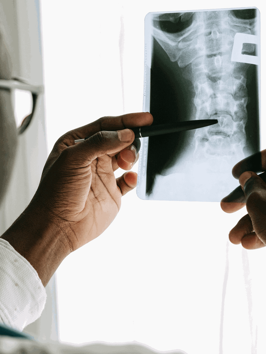
Assessment and differential diagnosis
Clinical interpretation reviews whether pain is axial or radicular, whether neurological findings are present and whether the MRI level plausibly matches the presentation. Routine MRI is not required for uncomplicated low back pain. Discography is invasive, controversial and not used as a routine confirmation test. Alternative sources such as facet joints, hip, sacroiliac joint and persistent pain mechanisms must be considered.
| Clinical pattern | Possible interpretation | Why it matters |
|---|---|---|
| Incidental fissure without concordant symptoms | No fissure-specific treatment required | Imaging abnormalities may be asymptomatic. |
| Axial pain without neurological deficit | Conservative care and functional monitoring | The exact pain generator may remain uncertain. |
| Fissure with disc herniation and radiculopathy | Treat the clinically relevant nerve-root condition | Neurological findings drive urgency. |
| Progressive weakness or cauda equina signs | Urgent assessment | The fissure label is not the priority. |

Treatment and decision-making
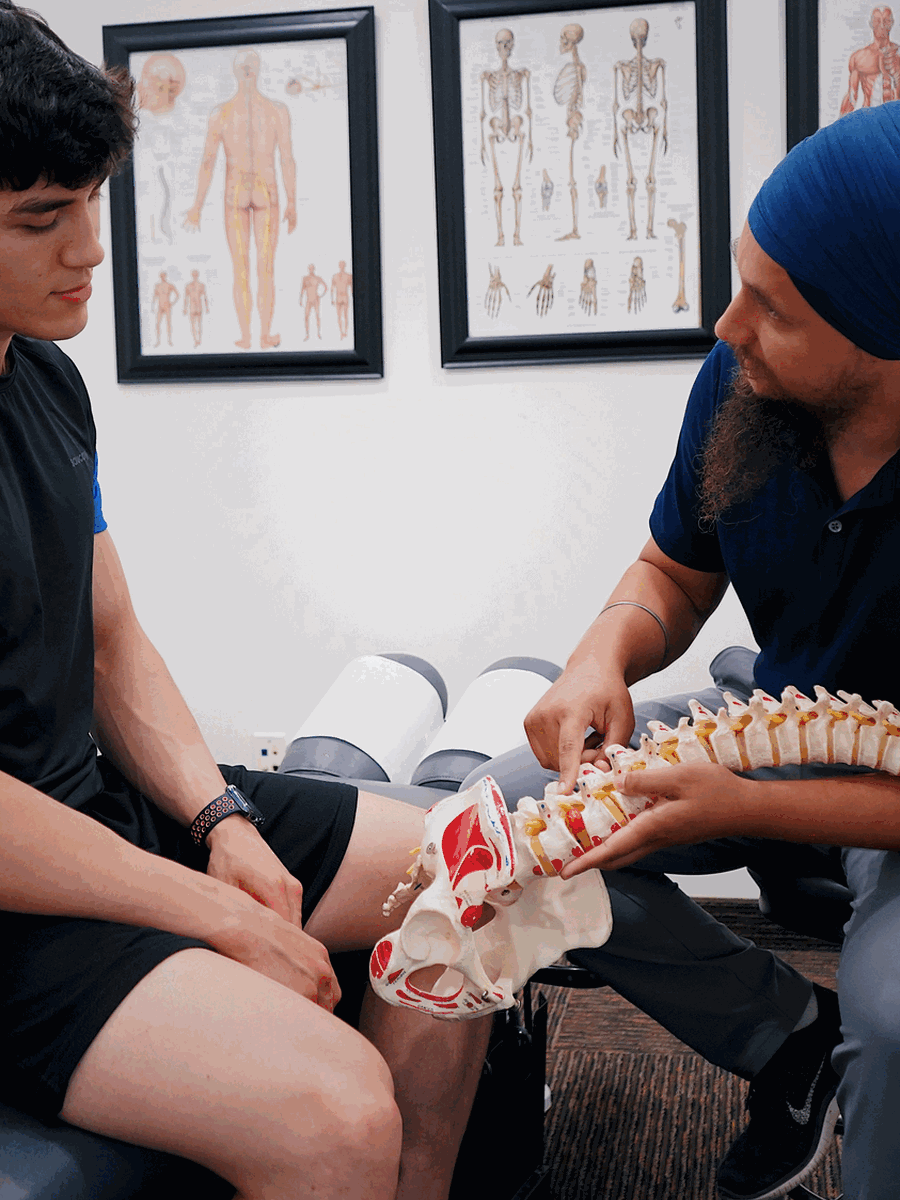
Management generally follows the broader low-back-pain or disc-herniation pathway: education, tolerable activity, exercise, symptom management and reassessment. Injections or surgery are not selected solely because an annular fissure appears on MRI. Claims that a technology “seals” or permanently repairs the annulus require direct evidence for the specific product and patient population.
Questions before choosing care
What is the leading diagnosis?
Ask which findings support it and which alternatives remain.
What test would change care?
Avoid testing that cannot alter the next step.
What result matters?
Track function, neurological safety and meaningful activity.
When should the plan change?
Define referral, stopping and escalation criteria.

Relevant specialized resources
Some resources may share ownership or editorial direction; this relationship is disclosed and the links are included for contextual relevance.
SOS Herniated Disc
Specialized English-language information about disc morphology and symptoms.
Frequently asked questions
Is an annular fissure the same as a herniated disc?
No. A fissure may exist without displaced disc material.
Does “tear” prove trauma?
No. The preferred term is fissure because the cause and age are often unknown.
Can a fissure be painless?
Yes.
What is a high-intensity zone?
A bright MRI signal in the annulus on certain T2-weighted images.
Does MRI show when the fissure occurred?
Usually not precisely.
Does every fissure need treatment?
No.
Can exercise worsen a fissure?
Exercise should be guided by symptoms and neurological safety rather than the label alone.
Can a fissure heal?
Disc tissue may change over time, but symptom recovery does not require complete MRI normalization.
Does spinal decompression seal the annulus?
This is not established as a general clinical outcome.
Does The Spine Page diagnose discogenic pain?
No.
Sources consulted
- Fardon et al. — Lumbar disc nomenclature version 2.0
- Fardon et al. — Standardized lumbar disc terminology
- American College of Radiology — Low Back Pain
Last editorial review: July 2026. General educational information only.
Dr. Sylvain Desforges, B.Sc., D.O., N.D., osteopath — The Spine Page
Continue exploring spine health
Use the condition and treatment hubs to compare related guides.
The Spine Page — The best treatments for your spinal problems — www.thespinepage.com