





L4-L5 or L5-S1 Sciatica in Montreal: Understanding Level and Nerve Root
The terms L4-L5 sciatica and L5-S1 sciatica are often used to describe an imaging level, but the disc level and the symptomatic nerve root are not always the same.
In Montreal as elsewhere, interpretation should connect side, pain distribution, sensation, strength, reflexes and imaging. An L4-L5 herniation may irritate the L5 root, while an L5-S1 lesion may affect S1.
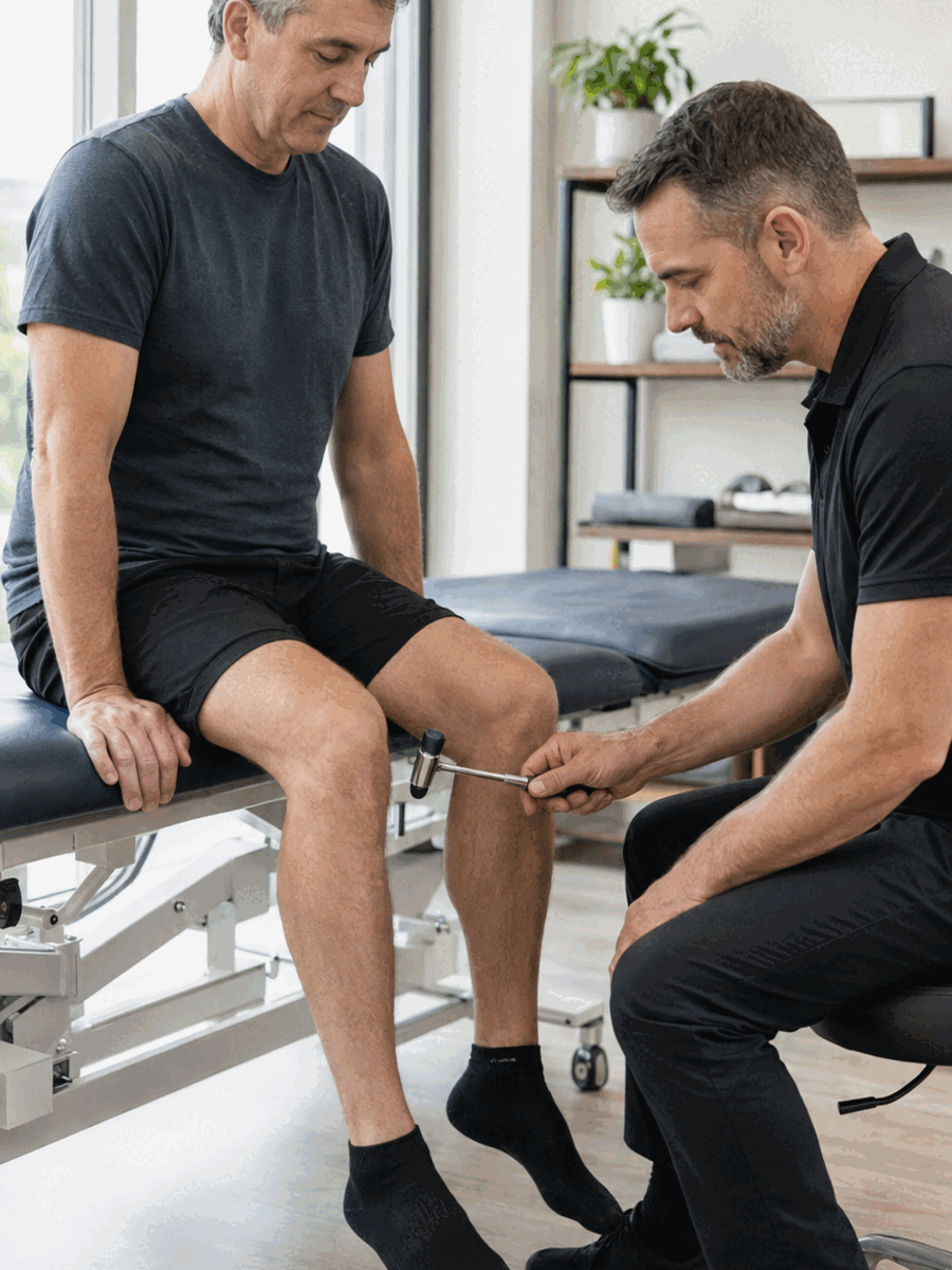
Do not confuse the spinal level with the nerve root
Dermatome and myotome maps are guides, not absolute boundaries. One patient may have overlapping findings, and imaging may show several abnormalities while only one is clinically concordant.
Connect L4-L5 with L5 and L5-S1 with S1, then assess strength, sensation, reflexes and function.
Clinical profile
Connect onset, distribution and modifying factors.
Neurology
Document strength, sensation, reflexes and gait.
Participation
Measure sleep, work, driving and meaningful activities.
Reassessment
Define when to continue, modify or refer.
Clinical, functional and neurological assessment
History should define onset, duration, side, aggravating positions, relieving factors, previous treatment and relevant medical or surgical history. Examination may compare mobility, neural tension, strength, sensation, reflexes and gait. A simple reproducible measure is more useful than a vague impression of feeling better.
Connect L4-L5 with L5 and L5-S1 with S1, then assess strength, sensation, reflexes and function. Imaging does not replace examination and is not routine; it is most useful when it can change the decision.
| Clinical situation | Possible step | Why |
|---|---|---|
| L4-L5 lesion affecting L5 | Lateral leg or top-of-foot pain; difficulty lifting the foot or great toe | Distribution and strength should be compared with imaging. |
| L5-S1 lesion affecting S1 | Posterior calf or outer-foot pain; difficulty rising on the toes | The Achilles reflex and plantar flexion can contribute to assessment. |
| Several abnormal MRI levels | Identify the most concordant level | Not every imaging abnormality is symptomatic. |

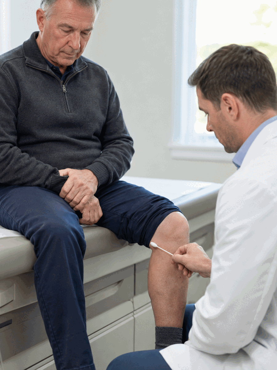
Build a measurable and revisable plan
The pathway may combine education, adjusted activity, progressive exercise, selected manual care and medical interventions when justified. Coherence matters more than accumulating techniques. Each option should answer a specific clinical question and include a functional goal the patient can understand.
Dermatome and myotome maps are guides, not absolute boundaries. One patient may have overlapping findings, and imaging may show several abnormalities while only one is clinically concordant. Expected benefits, limitations, risks and the reassessment schedule should be explained before starting.
Information to document before and during care
A credible pathway turns observations into verifiable goals and explicit progression criteria.
- Onset, duration, side and symptom distribution.
- Aggravating and relieving factors and positional tolerance.
- Strength, sensation, reflexes, gait and balance.
- Agreement among symptoms, function and imaging when available.
- Functional goals: sleep, walking, work, driving or daily activities.
- Stopping, escalation and urgent referral signs.
Follow function rather than a single number
Follow-up should not rely only on pain intensity. It should also monitor symptom distribution, strength, sensation, sleep quality, sitting tolerance, walking, work and meaningful activities. Neurological deterioration requires faster review than ordinary pain fluctuations.
Centralization of pain toward the back, improved walking or more stable strength may be encouraging. Conversely, more distal pain, expanding numbness or loss of capacity requires review.
Common mistakes to avoid
- Calling every leg pain pattern L5-S1 sciatica.
- Assuming the imaging level automatically identifies the root.
- Ignoring great-toe, foot or calf strength.
- Treating several MRI abnormalities without ranking concordance.
- Forgetting that hip, neuropathic or vascular conditions can mimic radiculopathy.
Questions to ask before starting
What is the working diagnosis?
Ask what supports it and which other causes remain possible.
What outcome will be measured?
Pain alone is insufficient; function and neurology complete follow-up.
When will the plan be reviewed?
Set a date and concrete criteria to continue, adjust or stop.
What are the referral thresholds?
Clarify signs requiring imaging, medical review or emergency care.

Related guides in the Montreal sciatica silo
Sciatica assessment in Montreal
Explore this clinical intent and its role in the pathway.
Spinal decompression and sciatica
Explore this clinical intent and its role in the pathway.
Disclosed clinical resource
Readers who wish to verify a local service may consult this separate, disclosed resource. Transactional information should be confirmed directly with the clinic.
TAGMED Clinic — Mount Royal / Montreal
Information about assessment and selected non-surgical care for pain compatible with sciatica. Address: 1140 Beaumont Avenue, Mount Royal, Quebec, H3P 3E5.
Frequently asked questions
Is sciatica always caused by a herniated disc?
No. Herniation is common, but foraminal stenosis, spinal stenosis, arthritis, spondylolisthesis or another condition can produce a radicular pattern.
Is MRI always required before treatment?
No. Imaging is most useful when it can change management, when a neurological deficit is present, when symptoms persist or when another cause must be clarified.
Does pain have to reach the foot?
No. Symptoms may stop in the buttock, thigh or calf. Distribution helps guide assessment but does not confirm the affected root by itself.
Is walking always recommended?
Walking is often useful when tolerated, but it may aggravate some patterns. Monitor whether pain travels farther, numbness increases or function declines.
When is sciatica an emergency?
Loss of bladder or bowel control, saddle anaesthesia, major or rapidly progressive weakness, fever with spinal pain or serious trauma requires urgent assessment.
Can treatment be selected from an X-ray alone?
X-rays mainly show bone, alignment and some degenerative changes. They do not directly show nerve roots or discs as MRI does.
How long should an approach be tried?
Timing depends on diagnosis, severity and the plan. A credible trial defines baseline measures, a reassessment point and criteria to continue, modify or stop.
Does less pain mean the nerve has recovered?
Not necessarily. Strength, sensation, gait, sleep and participation in activities should also be followed.
Does The Spine Page directly provide treatment in Montreal?
No. The Spine Page is a separate educational publication. The external clinical resource is disclosed and should be contacted directly for service information.
Which professional should be consulted?
The choice depends on symptoms and urgency. A clinician able to assess neurology, differential diagnoses and imaging indications can guide a safer pathway.
Main clinical sources
- NICE — Low back pain and sciatica in over 16s
- American College of Radiology — Low Back Pain
- World Health Organization — non-surgical management of chronic primary low back pain
- Cochrane — Traction for low-back pain
Last editorial review: August 2026. General educational information only.
Dr. Sylvain Desforges, B.Sc., D.O., N.D., osteopath – The Spine Page
Continue your research on sciatica in Montreal
Compare this page with the general guide, assessment pages and treatment options to maintain a coherent overview.
Dr. Sylvain Desforges, B.Sc., D.O., N.D., osteopath