


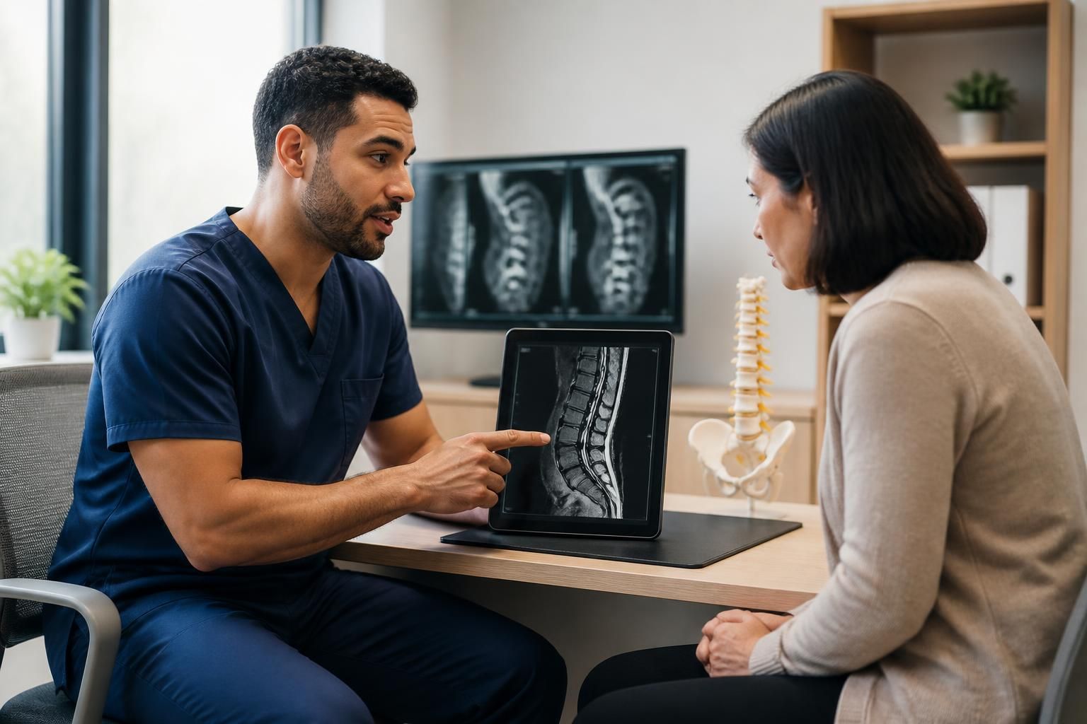


The use of MRI is a key tool in diagnosing sciatica, particularly in Rosemont–La Petite-Patrie, where access to quality medical imaging influences care. This examination detects structural changes in the sciatic nerve and its surrounding environment, thereby going beyond the traditional limitations of lumbar CT scans. It is particularly indicated when low back pain radiating into the leg persists without an obvious cause on conventional clinical assessment.
Understanding the contribution of MRI in diagnosing lumbosciatica involves considering the complexity of the sciatic nerve and its associated conditions. In Rosemont–La Petite-Patrie, the diagnostic approach emphasizes clinical assessment supported by targeted imaging, which is essential for guiding appropriate treatment, especially in atypical situations or those resistant to conservative approaches. These imaging techniques support clinicians in better identifying lesions and their mechanisms.
In brief: key points about MRI and diagnosing sciatica in Rosemont–La Petite-Patrie
- MRI provides detailed visualization of soft tissues and of the sciatic nerve along its entire course.
- It complements the clinical diagnosis to better differentiate the possible causes of lumbosciatica.
- The 2026 indications recommend MRI in cases of persistent symptoms, neurological deficits, or suspected complex conditions.
- CT remains preferred for rapid analysis of bone structures, but MRI provides essential precision for peripheral nerve lesions.
- In Rosemont–La Petite-Patrie, access to advanced imaging services facilitates this personalized assessment, which is essential for appropriate care.
Differences and complementarity between MRI and CT for diagnosing sciatica
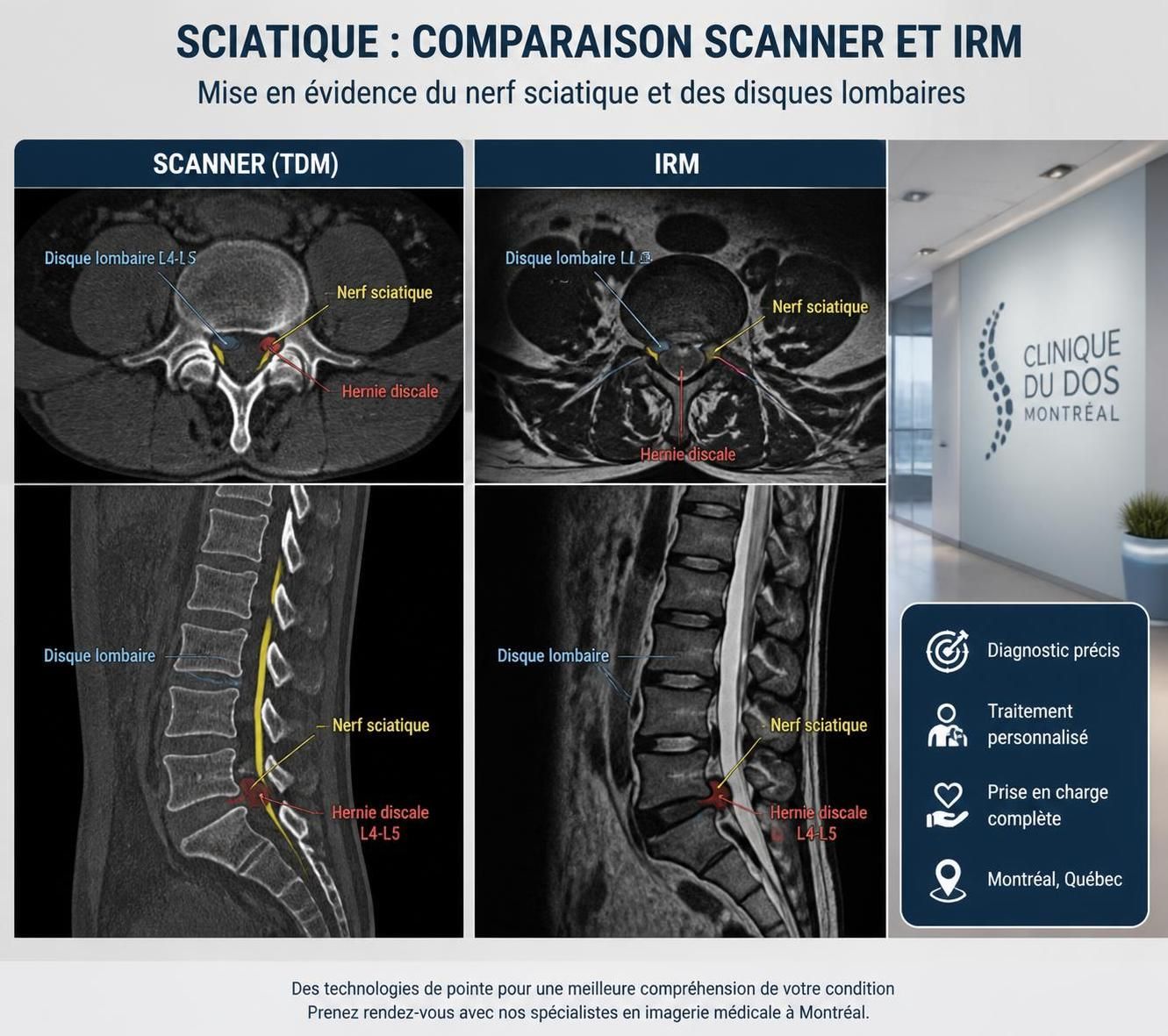
For a patient in Rosemont–La Petite-Patrie with pain consistent with sciatica, the choice of imaging examination often depends on the initial clinical assessment. CT, because of its short examination time and availability, is frequently used as a first-line option, especially to rapidly investigate bone abnormalities such as a vertebral fracture, osteophytosis, or osteoarthritis of the facet joints.
By contrast, MRI, which uses a magnetic field and radio waves, provides much more detailed visualization of soft tissues, including the intervertebral discs, nerve roots, sciatic nerve, and surrounding muscles. This is particularly important when disc protrusion, herniation, or nerve inflammation is suspected. The absence of radiation and superior image quality also make MRI more appropriate for sensitive populations, such as younger patients or pregnant women.
| Criterion | CT scan | MRI |
|---|---|---|
| Principle | X-rays | Magnetic field and radio waves |
| Examination duration | 5 to 20 minutes | 30 to 60 minutes |
| Primary visualization | Bones and dense structures | Soft tissues, nerves, intervertebral discs |
| Risks | Radiation exposure | No radiation |
| Indications for sciatica | Bone conditions, fractures | Nerve compression, inflammation, disc herniations |
This technical distinction is significant, particularly for understanding why MRI is preferred when sciatic pain persists or progresses despite initial care. Indeed, it is common for CT not to reveal a clear cause, especially when the lesion is located in soft tissues or within the complex structure of the sciatic nerve itself.
Specific role of sciatic nerve MR neurography in assessment in Rosemont–La Petite-Patrie
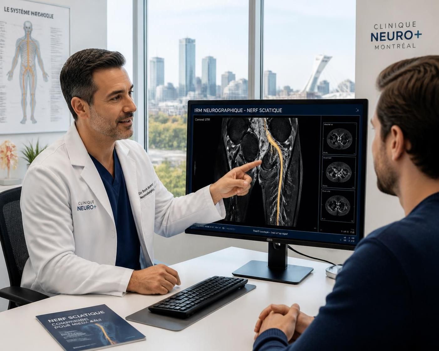
MR neurography is a specialized technique that provides targeted images not only of the spine, but also of the sciatic nerve along its entire course, from the pelvis to the leg. For patients in Rosemont–La Petite-Patrie with lumbosciatica and persistent or atypical symptoms, this technique often provides important additional information.
This method highlights signal variations that may indicate swelling, inflammation, compression, or nerve injury. It can also detect secondary involvement, such as muscle changes related to chronic nerve damage. This level of precision usefully complements neurological assessment and electrophysiological testing. In this regard, MR neurography is particularly recommended when clinical examination identifies motor deficits, numbness, or unexplained pain extending beyond the typical lumbar nerve roots.
For example, a patient may experience pain resembling sciatica, without CT or standard lumbar MRI findings accounting for the severity of the symptoms. In this context, targeted nerve imaging can help guide specific care, whether this involves neurovertebral decompression, personalized physiotherapy, or, in more complex cases, a neurosurgical consultation.
Specialists in the region, such as those at TAGMED Clinic in Mont-Royal, recommend this approach to optimize the initial assessment and adapt treatments according to the specific lesions identified.
Factors to consider when ordering an MRI to diagnose sciatica
The decision to order an MRI for lumbosciatica always requires an in-depth clinical analysis. In Rosemont–La Petite-Patrie, as elsewhere, national and international recommendations for 2025 emphasize caution in the use of imaging. MRI is not routinely used as a first-line examination, especially for recent pain without severe neurological signs.
- Duration and progression of symptoms : sciatic pain persisting beyond 6 weeks despite conservative treatment often supports ordering an MRI.
- Presence of neurological signs : progressive muscle weakness, numbness, and motor problems warrant a thorough MRI examination.
- Atypical or unexplained symptoms : when a lumbar origin does not appear evident, specialized imaging is indicated.
- Trauma or surgical history : these may require targeted MR neurography to assess peripheral nerve lesions or postoperative complications.
- Contraindications and limitations : claustrophobic patients, incompatible metal implants, or other constraints may lead to other imaging modalities being considered.
This rigorous selection aims to avoid unnecessary or counterproductive use of MRI while ensuring optimal investigation in complex or urgent cases. To explore the indications according to your situation, the recommended guide on medical examinations for sciatica provides relevant and up-to-date information.
The role of MRI in the care pathway and treatment options in Rosemont–La Petite-Patrie
Once the condition has been identified through clinical examination and, if necessary, supplemented by specialized MRI, care is guided by the causes and severity of the lesions. MRI can often confirm a disc herniation, foraminal stenosis, or nerve trauma, which are key elements in developing an appropriate treatment plan.
Recommended approaches include:
- Maintaining or gradually resuming appropriate physical activities, with careful monitoring of pain intensity.
- Tailored physiotherapy exercises targeting the spinal stabilizing muscles, often guided by the MRI diagnosis.
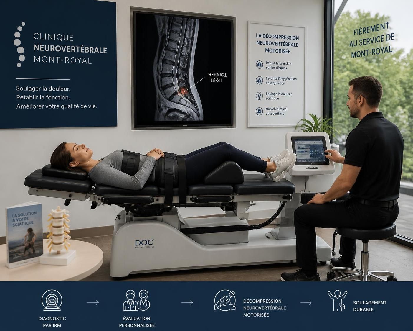
- Motorized neurovertebral decompression, offered at a clinic in Mont-Royal, particularly when MRI confirms a disc protrusion or nerve compression compatible with this treatment. This technique uses controlled traction intended to reduce pressure on the nerve root while respecting the patient’s anatomy.
- In cases of a significant neurological deficit, a specialized neurosurgical consultation may be required when imaging reveals severe compression or a lesion requiring intervention.
MRI is therefore only the beginning of a thoughtful diagnostic and therapeutic process that combines radiological analysis with detailed clinical assessment, in order to offer every patient in Rosemont–La Petite-Patrie the best possible care while avoiding redundant examinations and inappropriate treatments.
List: criteria for appropriate MRI ordering in the diagnosis of sciatica
- Symptoms persisting for more than 6 weeks despite conservative treatment.
- Onset or worsening of motor or sensory deficits.
- Atypical manifestations that do not correspond to typical lumbar radiculopathy.
- History of trauma, surgery, or local interventions in the pelvic area.
- Investigation for rare conditions such as tumours or inflammatory sequelae.
Table: Main clinical indications for ordering an MRI for a patient with sciatic pain in Rosemont–La Petite-Patrie
| Clinical situation | Why MRI is recommended | Possible treatment actions |
|---|---|---|
| Persistent unexplained sciatic pain | Identify a peripheral nerve lesion or disc herniation | Adapted exercises, physiotherapy, neurovertebral decompression |
| Progressive motor or sensory deficit | Identify significant nerve compression or a serious lesion | Neurosurgical consultation, intensive follow-up |
| Pelvic trauma or recent surgery | Assess nerve integrity and adjacent soft tissues | Conservative treatment or targeted surgical intervention |
| Atypical symptoms or unexplained pain | Differentiate among various conditions, tumours, or inflammation | Adapt treatment according to the identified cause |
Is MRI recommended for all low back pain radiating into the leg?
No. MRI is not routinely used for acute low back pain without warning signs. It is reserved for persistent, atypical cases or cases involving neurological deficits.
What are the advantages of sciatic nerve MR neurography?
It provides detailed visualization of the sciatic nerve and its surroundings, helping to precisely locate nerve lesions, which may affect treatment decisions.
How does MRI compare with CT for sciatica?
CT is fast and identifies bone abnormalities, whereas MRI provides detailed assessment of soft tissues and the nerve, offering an essential complementary evaluation in some cases.
Can MRI determine whether surgery is necessary?
MRI helps show the severity and location of nerve lesions, but the surgical decision also depends on clinical examination, symptom progression, and other tests.
Does MRI eliminate the need for other examinations?
No. MRI complements clinical examination and electrophysiological testing. Combining them provides the best approach to an accurate diagnosis.
If you experience persistent or atypical pain travelling down the leg, a thorough assessment in Rosemont–La Petite-Patrie may help clarify the likely cause. An approach combining clinical analysis, MRI imaging, and personalized advice remains essential for an appropriate care pathway.
To discuss your situation with specialists experienced in treating sciatica, feel free to consult the page dedicated to sciatica treatment options in Montréal or contact TAGMED Clinic.
Dr. Sylvain Desforges, B.Sc., D.O., N.D., osteopath
This content is intended for educational purposes and does not replace a personalized consultation.
