





Degenerative disc disease: what imaging changes mean and how symptoms are treated
Disc degeneration describes changes such as lower water content, reduced disc height, fissures and altered endplates. These findings are common and do not automatically identify the source of pain.
The task is to determine whether the changes plausibly relate to local pain, nerve-root symptoms or stenosis and whether another explanation is more likely.
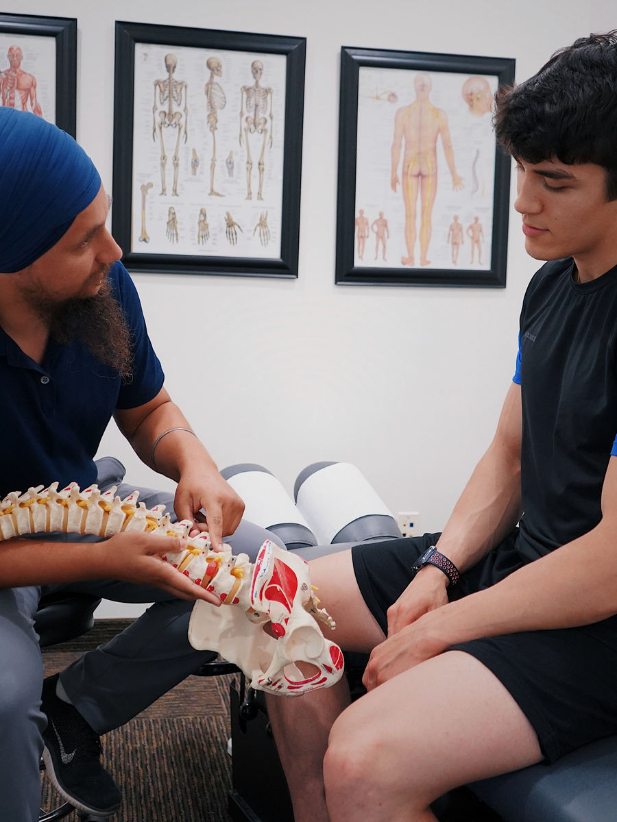
How disc changes may present
Some people have local axial pain; others develop radicular pain when a nerve root is irritated. Degeneration may coexist with facet pain or persistent pain mechanisms.
Movement and loading responses provide clues, but no single test proves a painful disc.
| Presentation | Possible mechanism | Caution |
|---|---|---|
| Local back or neck pain | Load-sensitive disc, endplate or adjacent joints. | Pain location alone cannot isolate the disc. |
| Radiating limb pain | Associated herniation or foraminal narrowing. | Neurological correlation is required. |
| Persistent multifactorial pain | Interaction of physical, sleep, stress and behavioural factors. | Do not focus only on MRI. |

Connect the image to the person
The assessment reviews onset, duration, activities, sleep, work, neurological symptoms and prior care. Examination considers motion, strength, sensation, reflexes and function.
Imaging is useful when serious pathology is suspected, deficits are present, symptoms persist or an invasive procedure is considered.
Clinical correlation
The level and side should match symptoms.
Functional baseline
Walking, sitting, lifting, sleep and work provide measurable targets.
Risk screening
Trauma, cancer, fever, osteoporosis and neurological change alter the pathway.

Treatment focuses on function
Guidance for persistent low back pain emphasizes education, exercise and person-centred combinations rather than one passive treatment.
When degeneration contributes to radicular pain or stenosis, medication, injections, decompression technology or surgery may be discussed according to diagnosis and deficit.
- Maintain tolerable activity instead of prolonged rest.
- Use graded strength and endurance training.
- Address sleep and fear of movement.
- Set measurable review points for passive care.
What changes the treatment decision?
A deficit or clearly correlated nerve compression matters more than the word “degeneration.”
A credible plan explains the target, timeline, alternatives and escalation criteria.
| Finding | Interpretation | Possible next step |
|---|---|---|
| Degeneration without neurological signs | Common age-related change. | Education, activity and rehabilitation. |
| Height loss with foraminal symptoms | Possible exiting-root irritation. | Neurological examination and targeted review. |
| Progressive deficit with compression | Higher-risk neurological presentation. | Prompt specialist assessment. |

Questions to ask before choosing care
Before beginning care for degenerative disc disease, ask the provider to explain the working diagnosis and the findings that support it. The discussion should distinguish a structural description from the clinical syndrome, clarify whether neurological function is stable and identify the specific outcome the treatment is intended to improve.
A high-quality plan also explains the expected timeline, reasonable alternatives, possible harms, cost and the criteria for changing course. No treatment should continue indefinitely because it produced a brief change in pain. The decision should be reviewed against measurable function and safety.
What is the target?
Ask which structure, mechanism or functional limitation the treatment is intended to address.
What result is realistic?
Clarify the expected change in pain distribution, walking, sleep, work or daily activity.
What are the alternatives?
Compare education, exercise, medication, procedures, technology-assisted care and surgery when relevant.
When will the plan be reviewed?
Set an early review point and define the findings that would stop, modify or escalate care.
How to monitor progress
Progress should be tracked with more than a pain score. Useful measures include how far symptoms travel, strength, sensation, walking tolerance, sleep, medication use and the ability to perform meaningful activities. Neurological change should always be documented separately from pain relief.
Temporary fluctuations are common. The important question is whether the overall trajectory is safer and more functional. New weakness, expanding numbness, altered bladder or bowel function or a substantial loss of walking ability should trigger reassessment rather than automatic continuation of the same treatment.
Continue with specialized resources
The Spine Page is an educational blog. The links below lead to independent specialized websites.
Clinique TAGMED — spinal decompression
Service-specific information about motorized spinal decompression and disc-related conditions.
SOS Sciatique — nerve-root symptoms
Focused guides about sciatica, warning signs and treatment questions.
Frequently asked questions
Is disc degeneration part of ageing?
Often, but clinical importance varies.
Does a dark disc prove pain?
No.
Can disc height be restored?
Treatment usually targets symptoms and function.
Is exercise safe?
Usually, with adaptation.
Does degeneration always progress?
Symptoms and imaging may not progress together.
Can it cause sciatica?
It may contribute through herniation or narrowing.
When is surgery considered?
When a treatable lesion matches severe persistent symptoms or progressive deficit.
Can decompression be considered?
Sometimes for selected presentations, with screening.
Must lifting be avoided forever?
Usually no.
Can The Spine Page interpret my MRI?
No.
Sources consulted
- World Health Organization — Guideline for non-surgical management of chronic primary low back pain
- Brinjikji et al. — Imaging features of spinal degeneration in asymptomatic populations
- Lumbar Disc Nomenclature: Version 2.0
- NICE — Low back pain and sciatica in over 16s
Last editorial review: July 2026. This page provides general education and does not replace individualized medical assessment.
Editorial review : Dr. Sylvain Desforges, B.Sc., D.O., N.D., osteopath — The Spine Page
Understand related disc problems
Compare disc degeneration, herniation and decompression questions.
The Spine Page — The best treatments for your spinal problems — www.thespinepage.com
Topic index: Disc degeneration and disc-space narrowing
Explore this topic cluster by search intent. Each link leads to a distinct guide directly related to the subject.