





Adjacent segment disease after spinal fusion: degeneration, symptoms and treatment
Adjacent segment degeneration is a radiographic change next to a fused spinal level. Adjacent segment disease means the neighbouring level becomes clinically symptomatic. The two terms should not be used interchangeably.
Fusion eliminates motion at one or more levels and can alter loading at neighbouring segments. At the same time, the adjacent discs and facets may already be ageing naturally or degenerating before surgery. New disc herniation, stenosis, instability, spondylolisthesis or deformity can develop above or below the fusion. A postoperative MRI change alone is not clinical disease unless it explains new symptoms and function loss.
Assessment and differential diagnosis
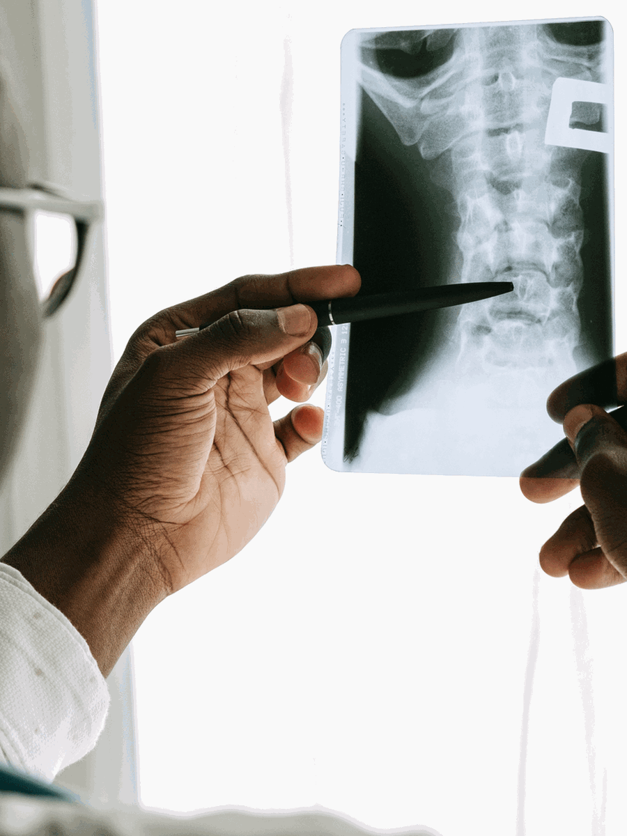
Evaluation reconstructs the original diagnosis, fusion levels, alignment, previous symptoms and symptom-free interval. The examination identifies whether new pain is axial, radicular, claudicant or myelopathic. Standing radiographs assess alignment, hardware, listhesis and disc-height change. Flexion–extension imaging may evaluate instability. MRI assesses discs, stenosis, neural compression and adjacent soft tissues. CT evaluates fusion integrity, hardware and bone. The differential includes nonunion, hardware problems, infection, sacroiliac or hip pain and persistent spinal pain syndrome.
| Clinical pattern | Possible next step | Why it matters |
|---|---|---|
| Radiographic change without new symptoms | Observation and risk-factor management | Not every adjacent change is disease. |
| New adjacent-level radiculopathy or claudication | Conservative care and targeted evaluation | The new level must explain the clinical pattern. |
| Instability or deformity with severe disability | Revision surgery may be considered | Decompression alone may not address instability. |
| Hardware failure, nonunion or infection | Different postoperative pathway | Do not label every postoperative problem as adjacent disease. |

Treatment and decision-making
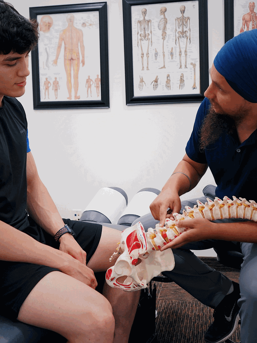
Stable symptoms may begin with education, activity modification, exercise, medication and selected injections. Treatment should address the actual new lesion rather than the fact of prior fusion. Surgical options include decompression alone in selected stable stenosis, extension of the fusion, interbody fusion or motion-preserving strategies in carefully selected cases. Alignment, bone quality, previous fusion length, paraspinal muscle condition and the need for further facet removal influence the revision plan.
How to interpret the finding in practice
The key question is whether the neighbouring level is newly symptomatic and mechanically or neurologically responsible. Preventing all future degeneration is impossible, but surgical planning can reduce avoidable risk.
Compare old imaging
Pre-existing adjacent degeneration changes the interpretation.
Check fusion integrity
Nonunion can imitate adjacent disease.
Assess global alignment
Sagittal imbalance may increase adjacent loading.
Limit assumptions
New back pain may arise from the hip, SI joint or muscles.
Preserve healthy structures
Revision planning should minimize unnecessary facet and muscle injury.
Discuss future levels
Extending a fusion may create another adjacent segment.
How to monitor progress and avoid treatment errors
A credible treatment plan should define what improvement means before care begins. Pain intensity alone is not enough, because pain may fluctuate while neurological function, walking, sleep or daily activity changes in another direction. The diagnosis should also be reconsidered when the expected response does not occur.
Establish a baseline
Record symptom distribution, strength, sensation, walking, sleep, medication use and the activities that matter most.
Use a planned review point
Decide when progress will be evaluated rather than continuing automatically until a package is completed.
Separate relief from diagnosis
Temporary improvement after an injection, manual treatment or device does not prove that the proposed structure was the true cause.
Watch the neurological trajectory
New weakness, spreading numbness or balance change matters even if pain has decreased.
Revisit competing diagnoses
Hip, sacroiliac, vascular, inflammatory and postoperative causes may overlap with the initial explanation.
Escalate proportionately
Imaging, specialist referral or surgery should be considered when the risk or functional loss justifies it.
Common mistakes to avoid
- Treating the largest imaging abnormality rather than the clinically relevant lesion.
- Assuming that a diagnostic label remains correct when the symptom pattern changes.
- Repeating passive care despite no measurable functional progress.
- Using one negative test to exclude a condition that remains clinically plausible.
- Delaying urgent assessment to complete a predetermined treatment schedule.
- Generalizing results from a narrowly selected study population to every patient with a similar MRI term.
Shared decision-making is strongest when uncertainty is stated directly. A provider should explain what is confirmed, what remains probable, which alternatives still matter and what findings would change the plan. For complex or postoperative presentations, a second opinion from a clinician experienced with the specific condition may be more useful than adding another non-specific treatment.
Questions before choosing care
What is the leading diagnosis?
Ask which findings support it and which alternatives remain.
What test would change care?
Avoid testing that cannot alter the next step.
What result matters?
Track function, neurological safety and meaningful activity.
When should the plan change?
Define referral, stopping and escalation criteria.

Relevant specialized resources
Some resources may share ownership or editorial direction; this relationship is disclosed and the links are included for contextual relevance.
Frequently asked questions
Is adjacent degeneration the same as adjacent disease?
No.
How soon can adjacent disease appear?
It may appear years later, but timing varies.
Does fusion always cause adjacent disease?
No.
Can the level below the fusion be affected?
Yes.
Can exercise prevent it?
Exercise supports function but cannot guarantee prevention.
Does disc replacement prevent adjacent disease?
It may alter biomechanics, but long-term clinical superiority is not universal.
Is revision fusion always required?
No.
Can decompression alone be used?
Sometimes, when the adjacent segment is stable.
Can symptoms come from the SI joint after fusion?
Yes.
Does The Spine Page evaluate prior fusions?
No.
Sources consulted
- Huang et al. — Adjacent segment disease risk factors and treatment
- Cannizzaro et al. — Lumbar adjacent degeneration meta-analysis
- Mesregah et al. — Cervical adjacent disease risk factors
- McNamee et al. — Paraspinal musculature and adjacent disease
Last editorial review: July 2026. General educational information only.
Dr. Sylvain Desforges, B.Sc., D.O., N.D., osteopath — The Spine Page
Continue exploring spine health
Use the condition and treatment hubs to compare related guides.
The Spine Page — The best treatments for your spinal problems — www.thespinepage.com