

Group of muscles predominantly associated with a spinal nerve root, used as a guide during motor examination.
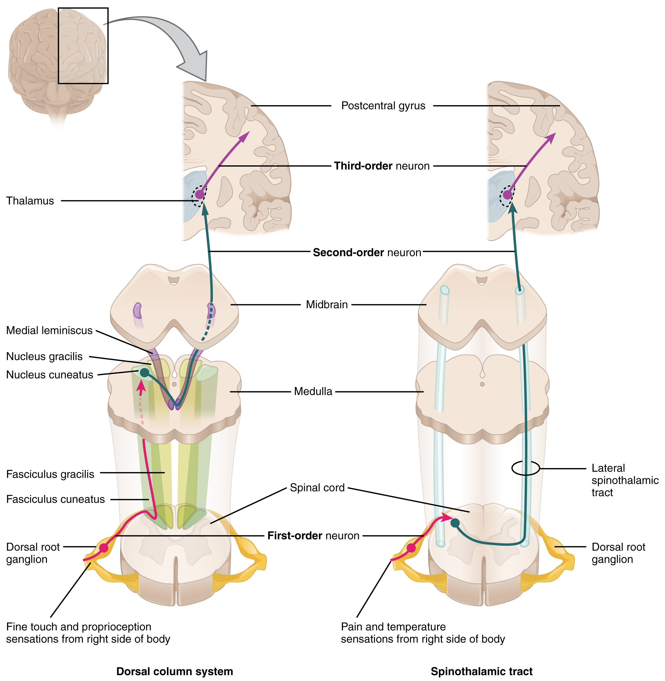
OpenStax College — CC BY 3.0. Image source.

Understanding the structure and its role
In neurological examination, a myotome describes a functional muscle group mainly associated with a spinal nerve level. Most muscles receive contributions from several roots, so tests are not on-off switches for a single root. Knee extension and lifting the foot are examples of functions examined.
Symptoms and assessment
The clinician compares sides, looks for fatigability and distinguishes neurological weakness from pain-related inhibition and deconditioning. Weakness consistent with sensory and reflex findings strengthens suspicion of root disease. Progressive weakness requires reassessment even if pain is improving.
Assessment considers symptom onset, distribution, restricted activities and medical history. Imaging findings must be compared with the clinical examination: a visible abnormality does not, on its own, establish the source of pain.
Management and outlook
Exercises are dosed to capacity and diagnosis. Forcing a markedly weak muscle without understanding the cause can delay useful referral. Recovery is documented through comparable tests, rather than only a sense of improved strength after a session.
For a mechanical problem without concerning findings, care aims for a gradual return to activity, individually selected exercises and measurable functional goals. Manual care may complement this approach when the examination supports it. Progress is judged through walking, sleep and daily tasks, as well as symptoms, rather than through an image alone.
When to seek prompt assessment
New or progressive weakness, newly impaired walking, loss of sensation around the saddle region, or new bladder or bowel control problems require urgent medical assessment.
Frequently asked questions
Are myotomes and dermatomes interchangeable?
No. The former concern motor function; the latter concern skin sensation.
Does one weak test prove a herniation?
No. Pain, tendon, muscle and peripheral nerve causes also need consideration.
Can imaging establish the diagnosis alone?
No. Findings must fit the clinical history and examination before they guide treatment.
Should all activity stop?
Usually not: loading is adjusted and gradually increased according to tolerance and the diagnosis.
How should progress be monitored?
Track functional abilities, neurological symptoms and resumed activities so care can be adjusted.
Concept diagram showing key relationships; this is not a scan or an anatomical drawing to scale.
Role of TAGMED care
At TAGMED, this information helps clarify your history, guide assessment and determine whether conservative care is appropriate or medical referral is needed. An anatomical structure, sign or test result is not, by itself, an indication for decompression, laser, shockwave or manual treatment. Care selection depends on the cause of symptoms and your clinical circumstances.
Medical references
- McGovern Medical School, UTHealth Houston. Anatomy of the Spinal Cord — Neuroscience Online.
- American Academy of Orthopaedic Surgeons. Cervical Radiculopathy (Pinched Nerve).
- MedlinePlus / A.D.A.M.. Femoral nerve dysfunction.
References document the topic; their inclusion does not imply endorsement of TAGMED services by those organizations.
Educational information from The Spine Page. This entry does not replace an individual assessment. Documentary version: October 12, 2026. Sylvain Desforges, B.Sc., D.O., N.D., osteopath and naturopath.
TAGMED clinic contact details
For non-urgent consultations and information about available services.
Clinique TAGMED — Terrebonne
1150, rue Lévis, Suite 200
Terrebonne (Québec) J6W 5S6
450‑704‑4447
Clinique TAGMED — Montréal
1140, avenue Beaumont
Mont-Royal (Québec) H3P 3E5
1‑877‑672‑9060